Warning: fopen(/home/virtual/colon/journal/upload/ip_log/ip_log_2025-03.txt): failed to open stream: Permission denied in /home/virtual/lib/view_data.php on line 95 Warning: fwrite() expects parameter 1 to be resource, boolean given in /home/virtual/lib/view_data.php on line 96 Clinical Characteristics of Colorectal Carcinoid Tumors
Skip Navigation
Skip to contents

Ann Coloproctol : Annals of Coloproctology

OPEN ACCESS
SEARCH
Search

Articles

Page Path
HOME > J Korean Soc Coloproctol > Volume 27(1); 2011 > Article
Original Article
Clinical Characteristics of Colorectal Carcinoid Tumors
1Colonoscopy Study Group of Korean Society of Coloproctology
Journal of the Korean Society of Coloproctology 2011;27(1):17-20.
DOI: https://doi.org/10.3393/jksc.2011.27.1.17
Published online: February 28, 2011

Colonoscopy Study Group of Korean Society of Coloproctology, Seoul, Korea.

Correspondence to: Jae-Bum Lee, M.D. Department of Surgery, Daehang Hospital, 481-10 Bangbae-dong, Seocho-gu, Seoul 137-820, Korea. Tel: +82-2-6388-8564, Fax: +82-2-6388-8673, commonlee@naver.com
• Received: April 2, 2010   • Accepted: August 25, 2010

© 2011 The Korean Society of Coloproctology

This is an open-access article distributed under the terms of the Creative Commons Attribution Non-Commercial License (http://creativecommons.org/licenses/by-nc/3.0) which permits unrestricted non-commercial use, distribution, and reproduction in any medium, provided the original work is properly cited.

prev next
  • 4,960 Views
  • 49 Download
  • 19 Crossref
  • 24 Scopus
  • Purpose
    The carcinoid tumor was recently categorized as a malignant disease due to its possibility of metastasis. This study was aimed to investigate the clinical characteristics and the metastatic rate of colorectal carcinoid tumors.
  • Methods
    Charts were reviewed for 502 patients diagnosed with and treated for colorectal carcinoid tumors between January 2006 and December 2009. The location, size, depth and metastatic status of the tumors were collected.
  • Results
    Including 24 synchronous tumors from 12 patients, 514 carcinoid tumors were removed. Most of them were found in the rectum (97.3%). The male-to-female ratio was 1.38 to 1, and mean age was 50.2 ± 11.4 years. The mean tumor size was 5.8 ± 3.6 mm. Less than 10-mm-sized tumors had a 1.95% lymph node metastatic rate; tumors with sizes from 10 mm to 20 mm and larger than 20 mm had 23.5% and 50% lymph node metastatic rates, respectively. Two cases had distant metastasis; one with a 22-mm-sized tumor metastasized to the liver, and the other with a 20-mm-sized tumor metastasized to the peritoneum. Among 414 patients who completed metastatic studies, 93.8% were classified as stage I, 0.9% as stage II, 4.8% as stage III and 0.5% as stage IV.
  • Conclusion
    Colorectal carcinoid tumors smaller than 10 mm have a low rate of lymph node metastasis, but those sized 10 mm or larger incur significant risk. Further investigation regarding additional risk factors should be done to develop proper treatment guidelines for these tumors.
Carcinoid tumors are the most common neuroendocrine tumor in the gastrointestinal system. The incidence of carcinoid tumors varies depending on the organ. However, the rectum and the ileum are the most prevalent tumor sites in the gastrointestinal system [1].
Among gastrointestinal carcinoid tumors, colonic carcinoids have been reported to be relatively rare while rectal carcinoid tumors have been reported to be the most prevalent [1-4]. Generally, carcinoid tumors have been recognized as having low malignant potential. However, recent studies have shown considerable rates of lymph node metastasis in carcinoid tumors. As well, the American Joint Council on Cancer (AJCC) has classified carcinoid tumors as malignant tumors [5].
The incidence of colorectal carcinoid tumors in Korea may be on the rise as the degree of screening colonoscopy increases [6, 7]. Although there have been reports of small-sized carcinoid tumors with lymph node metastasis, it is very difficult to assess national prevalence and rates of metastasis because previous domestic studies were limited to a single institute and had a limited volume of patients. Thus, the aim of this study is to investigate clinical characteristics focusing on the metastatic rate of colorectal carcinoid tumors through a large scale study.
The colonoscopy study group of the Korean Society of Coloproctology conducted a retrospective study of colorectal carcinoid tumors in collaboration with eight institutes from January 2006 to December 2009. We gathered data such as age, gender, site, size, invasion depth and the presence of metastasis through the collection and review of medical records.
Rectal carcinoids were defined as tumors located within 15 cm of the anal verge while tumors more than 15 cm above the anal verge were regarded as colonic carcinoids. The tumors were classified into three groups according to tumor size: 1) a tumor diameter less than 10 mm, 2) a 10-mm to 20-mm tumor diameter, and 3) a diameter larger than 20 mm. Tumor sizes were confirmed through pathology reports. In the case of tumor sizes not being clearly described in the pathology reports, the size was determined according to the colonoscopy reports. Treatment methods were classified as endoscopic resection, transanal resection and radical operation.
In cases treated by radical operation, the presence of lymph node metastasis was determined by using a pathology report while in cases treated by local resection, including colonoscopic removal, metastatic status were assessed by radiologic studies (computer tomography [CT] scan). Cases which did not undergo metastatic work ups were excluded from the analysis of lymph node metastasis. If there were synchronous carcinoid tumors, only the largest tumors were included in the analysis of lymph node metastasis.
The tumor disease stages were determined according to the AJCC Cancer Staging 7th edition, revised in 2010. Statistical analysis was done with the Fisher's exact test and a P-value less than 0.05 was considered to be statistically significant.
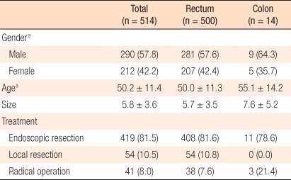
Five hundred two patients were treated for carcinoid tumors during the study period. In 12 patients, two carcinoid tumors were detected simultaneously (synchronous carcinoid tumor, 2.4%); thus, the total number of resected carcinoid tumors was 514. Fourteen carcinoid tumors were found in the colon, which accounts for 2.7%, and 500 carcinoid tumors (97.3%) were detected in the rectum. The ratio between males and females was 1.38:1, and the mean patient age was 50.2 years. The average tumor size was 5.8 mm, and a regional resection, including colonoscopic treatments, was performed in 473 cases (92%) (Table 1).
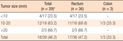
The result of pathological tests for 39 cases treated by radical surgery showed 18 cases of lymph node metastasis (46.2%). The relationship of lymph node metastasis to tumor size for tumors smaller than 10 mm was 23.5%, for tumors between 10 mm and 20 mm, it was 63.2%, and for tumors larger than 20 mm, it was was 66.7% (Table 2).
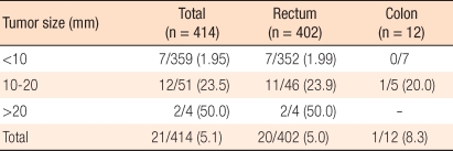
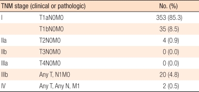
Metastasis was assessed through CT scans or pathological examinations in 414 cases. When cases showing metastasis findings on the test were considered as positive for lymph node metastasis, the aforementioned incidence according to tumor size for tumors smaller than 10 mm was 1.95% while for sizes between 10 mm and 20 mm, it was 23.5%, and for sizes larger than 20 mm, it was 50.0%. As tumor sizes increased, the metastasis incidence also increased (Table 3), and the frequency of lymph node metastasis in cases with carcinoid tumors smaller than 10 mm was confirmed to be significantly lower than in other groups (P < 0.001). Regarding two cases with distant metastasis, one rectal case had metastasis to the liver, and the other colonic case involved peritoneal metastasis. The result of the TNM disease stage classification according to the AJCC is shown in Table 4.
Generally, the incidence of carcinoid tumors of the colon has been reported to be very low and comprises 4-8% of carcinoid tumors in the digestive system [1-4]. Colonic carcinoid tumors are detected in the cecum primarily, and two-thirds of colonic carcinoid tumors have been reported to occur in the right side of the colon [8]. More than half of carcinoid tumors of the colon are associated with lymph node metastasis or distant metastasis at the time of detection. Thus, prognosis is poor, with 5-year survival rates reported to be approximately 40-70% [1, 9].
In our study, although it was limited to the colon and the rectum, the tumor incidence in the colon was 2.7%, which is lower than the generally accepted incidence rate. As well, the number of cases treated with radical surgery was only three. Among the three, one case had lymph node metastasis, and another case had peritoneal metastasis. For the remaining eleven cases, the tumors were resected locally through colonoscopy. Using these data, estimating the lymph node metastasis rate in terms of colonic carcinoid tumors is very difficult, making risk predictions infeasible.
The proportion of colonoscopic resections for colonic carcinoid tumors in our series was relatively high in relation to general treatment guidelines, which recommend radical surgery for all colonic carcinoids. A possible explanation for this is that contrary to the usual experience of finding colonic carcinoid tumors at advanced stages, many colonic tumors in our series may were found incidentally at smaller sizes during diagnosis, deeming colonoscopic resection appropriate.
Most carcinoid tumors we detected were in the rectum, correlating with known findings as to the organ in which carcinoid tumors are detected most frequently. In our study, cases involving rectal carcinoid tumors totaled 500, accounting for approximately 97.3%, with an average size of 5.7 mm. The frequency of lymph node metastasis for rectal carcinoid tumors was found to be 5.0%. Most rectal carcinoid tumors were treated by endoscopic or regional resection, such as transanal resection.
To evaluate the risk of metastasis, pathological examination of lymph nodes should be implemented. However, small rectal carcinoids are known to have little risk of metastasis, making local treatment desirable and reserving radical surgery for selected cases with risk factors associated with lymph node metastasis. Therefore, we substituted radiologic evaluation with CT scans for pathologic examination to confirm lymph node metastasis. Although CT scans do not reflect lymph node metastasis as accurately as a pathologic examination, if no sign of metastasis appears in the CT scan, we regard the case as negative for lymph node metastasis.
Four hundred four cases undergoing radical surgery and/or CT scan were included in the evaluation of metastasis. There was a direct correlation between tumor size and lymph node metastasis. The lymph node metastasis rate was approximately 2% if the tumor size was less than 10 mm and increased gradually up to 50% as the tumor size increased to larger than 20 mm. For tumor sizes between 10 mm and 20 mm, the risk of lymph node metastasis was as high as 24%, which was similar to the results reported by Soga et al. [2]. This is thought to be a very important finding in deciding treatment modality. A consensus exists for which treatment method is appropriate for rectal carcinoid tumors with sizes between 10 mm and 20 mm.
Carcinoid tumors develop in the mucosal gland and gradually infiltrate to the lamina propria, submucosal layer and deeper layer of the bowel wall. As the disease stage advances, carcinoid tumors may be accompanied by lymph node or distant metastasis. These clinical behaviors are the characteristics of malignant neoplasms. Thus, it is reasonable to regard colorectal carcinoids as malignant tumors. In diverse reports, lymph node metastasis and distant metastasis were reported and are thought to be tumors with a malignant potential comparable to that of an adenocarcinoma [2, 3]. In the recently revised AJCC cancer staging, carcinoid tumors are classified as a malignant disease. The stage classifications are based on the size of the tumor, the involved layer of the bowel wall and the presence or absence of lymph node or distant metastasis. According to this staging system, our series of cases are classified into 388 stage I cases, 4 stage IIA cases, no stage IIB or IIIA cases, 20 stage IIIB cases, and 2 stage IV cases. The staging results reflect that most cases were early lesions and were detected incidentally by screening colonoscopy.
In cases of rectal carcinoids, the risks of lymph node metastasis and distant metastasis vary depending on the tumor size, making tumor size the most important factor in deciding on a treatment method. The reported risk of lymph node metastasis for rectal carcinoids smaller than 10 mm is usually less than 3%; thus, regional treatment including colonoscopic resection is appropriate. However, radical surgery is recommended for tumors larger than 20 mm, where the risk of metastasis is known to be 60-80%. Treatment methods for tumors with sizes between 10 mm and 20 mm are controversial, and no established treatment guideline exists.
Known risk factors, other than tumor size, are tumor invasion beyond the proper muscle, the degree of cell differentiation and the mitotic rate. A population-based study in Japan reported 3.7% lymph node metastasis for rectal carcinoids with sizes of 5 mm and less and 10% for tumors with sizes less than 10 mm [2]. These results complicate deciding on treatment methods even in small-sized rectal carcinoids. Therefore, if lymph node metastasis is to be predicted accurately, additional studies on risk factors are required, and treatment methods should be determined considering various risk factors [9-11].
The large number of cases involved in this study aided in revealing meaningful data in terms of the risk of lymph node metastasis, yet, some limitations should be mentioned. First, it was a retrospective cross-sectional study and collected data were restricted to findings when tumors were detected and treated. Second, there may have been selection bias. In some institutions, only cases involving radical surgery were included. These cases may have had risk factors associated with lymph node metastasis. Third, we could not estimate the overall incidence, and this study lacked long-term follow-up results in terms of recurrence. This may play a role in underestimating the risk of lymph node metastasis. Additional studies on recurrence and the rate of metastasis through long-term follow-up are required in the future.
In rectal carcinoid tumors, the size of the tumor is directly related to the frequency of lymph node metastasis. In carcinoid tumors with sizes smaller than 10 mm, the risk of lymph node metastasis was low; thus, it could be treated by regional treatments including colonoscopic resection. However, in carcinoid tumors with sizes larger than 10 mm, the risk of lymph node metastasis increased remarkably. Especially, rectal carcinoid tumors with sizes between 10 mm and 20 mm showed a lymph node metastasis rate of 23.5%, requiring treatment to be determined by considering various risk factors. Additional studies on the risk factors for lymph node metastasis are needed.
We thank Dr. Hyun-Shig Kim, Dr. Hee-Cheol Jang (Seoul Song Do Hospital), Dr. Kyung-Su Han, Dr. Chang-Won Hong, Dr. Dae-Kyung Sohn (National Cancer Center), Prof. Won-Kyung Kang, Prof. Hye-Young Sung, Prof. Seong-Taek Oh (The Catholic University of Korea), Prof. Sung-Bum Kang, Prof. Duck-Woo Kim (Seoul National University Bundang Hospital), Prof. Hyoung-Chul Park, Prof. Bong-Hwa Lee (Hallym University College of Medicine), Dr. Seung-Kyu Jeong, Dr. Dong-Hyun Choi, Dr. Yong-Taek Ko (Yang Hospital), Dr. Eun-Jung Lee, Dr. Eui-Gon Youk (Daehang Hospital), Dr. Seok-Won Lim (Hang Clinic of Coloproctology) for their contribution to this study. They provided valuable data for this study.

No potential conflict of interest relevant to this article was reported.

  • 1. Modlin IM, Lye KD, Kidd M. A 5-decade analysis of 13,715 carcinoid tumors. Cancer 2003;97:934–959. ArticlePubMed
  • 2. Soga J. Early-stage carcinoids of the gastrointestinal tract: an analysis of 1914 reported cases. Cancer 2005;103:1587–1595. ArticlePubMed
  • 3. Konishi T, Watanabe T, Kishimoto J, Kotake K, Muto T, Nagawa H, et al. Prognosis and risk factors of metastasis in colorectal carcinoids: results of a nationwide registry over 15 years. Gut 2007;56:863–868. ArticlePubMedPMC
  • 4. Park YB, Kim JI, Ha BH, Cheung DY, Kim TH, Kim CH, et al. Endoscopic treatment of gastrointestinal carcinoid tumors. Korean J Med 2007;73:274–282.
  • 5. Edge SB. American Joint Committee on Cancer. AJCC cancer staging manual. 2010.7th ed. New York: Springer.
  • 6. Park JB, Cha JM, Lee JI, Choe JW, Joo KR, Jung SW, et al. A case of small rectal carcinoid tumor with local lymph node metastases. Korean J Gastrointest Endosc 2009;38:52–56.
  • 7. Chang JH, Kim SW, Chung WC, Kim YC, Jung CK, Paik CN, et al. Clinical review of gastrointestinal carcinoid tumor and analysis of the factors predicting metastasis. Korean J Gastroenterol 2007;50:19–25. PubMed
  • 8. Raut CP, Kulke MH, Glickman JN, Swanson RS, Ashley SW. Carcinoid tumors. Curr Probl Surg 2006;43:383–450. ArticlePubMed
  • 9. Maggard MA, O'Connell JB, Ko CY. Updated population-based review of carcinoid tumors. Ann Surg 2004;240:117–122. ArticlePubMedPMC
  • 10. Ramage JK, Goretzki PE, Manfredi R, Komminoth P, Ferone D, Hyrdel R, et al. Consensus guidelines for the management of patients with digestive neuroendocrine tumours: well-differentiated colon and rectum tumour/carcinoma. Neuroendocrinology 2008;87:31–39. ArticlePubMed
  • 11. Kim BN, Sohn DK, Hong CW, Han KS, Chang HJ, Jung KH, et al. Atypical endoscopic features can be associated with metastasis in rectal carcinoid tumors. Surg Endosc 2008;22:1992–1996. ArticlePubMed
Table 1
Clinical characteristics of colorectal carcinoid tumors
jksc-27-17-i001.jpg

Values are presented as number (%) or mean ± SD.

aTotal number of patients is 502.

Table 2
Rate of lymph node metastasis in patients who underwent radical surgery
jksc-27-17-i002.jpg

The values in parentheses are presented as percent.

aTwo synchronous tumors in two patients (total number of carcinoid tumors is 41).

Table 3
Rate of lymph node metastasis in 414 colorectal carcinoisa
jksc-27-17-i003.jpg

The values in parentheses are presented as percent.

aCases of radical operation or metastasis-evaluation using CT scan.

Table 4
Tumor-node-metastasis (TNM) staging of colorectal carcinoid tumors according to American Joint Council on Cancer, 7th edition (n = 414)
jksc-27-17-i004.jpg

T1, tumor invades lamina propria or submucosa and size ≤ 2 cm; T1a, tumor size < 1 cm in greatest dimension; T1b, tumor size 1 to 2 cm in greatest dimension; T2, tumor invades muscularis propria or size > 2 cm with invasion of lamina propria or submucosa; T3, tumor invades through the muscularis propria into the subserosa, or into non-peritonealized pericolic or perirectal tissues; T4, tumor invades peritoneum or other organs; N0, no regional lymph node metastasis; N1, regional lymph node metastasis; M0, no distant metastasis; M1, distant metastasis.

Figure & Data

References

    Citations

    Citations to this article as recorded by  
    • Clinical outcomes and risk factors associated with poor prognosis after endoscopic resection of 10–20 mm rectal neuroendocrine tumors: a multicenter, retrospective study of 10-year experience
      Ja Jun Goo, Dong Hoon Baek, Hyung Wook Kim, Hong Sub Lee, Jong Yoon Lee, Su Bum Park, Geun Am Song, Sang Heon Lee, Jong Hoon Lee
      Surgical Endoscopy.2023; 37(7): 5196.     CrossRef
    • Stratification of rate of lymph node metastasis according to risk factors and oncologic outcomes in patients who underwent radical resection for rectal neuroendocrine tumors
      Myung Jae Jin, Sung Sil Park, Dong-Eun Lee, Sung Chan Park, Dong Woon Lee, Kiho Yu, Hee Jin Chang, Chang Won Hong, Dae Kyung Sohn, Kyung Su Han, Bun Kim, Byung Chang Kim, Jae Hwan Oh
      Annals of Coloproctology.2023; 39(6): 467.     CrossRef
    • Risk factors of lymph node metastasis in rectal neuroendocrine tumors*
      Donghong Liang, Zhennan Niu, Xiaofang Sun, Changjuan Meng, Zhuang Liu
      Oncology and Translational Medicine.2022; 8(4): 186.     CrossRef
    • Risk of lymph node metastasis after endoscopic treatment for rectal NETs 10 mm or less
      Yutaka Inada, Naohisa Yoshida, Kohei Fukumoto, Ryohei Hirose, Ken Inoue, Osamu Dohi, Takaaki Murakami, Kiyoshi Ogiso, Akira Tomie, Munehiro Kugai, Hiroyuki Yoriki, Yoshikazu Inagaki, Daisuke Hasegawa, Kotaro Okuda, Takashi Okuda, Yukiko Morinaga, Mitsuo K
      International Journal of Colorectal Disease.2021; 36(3): 559.     CrossRef
    • Grade G2 Rectal Neuroendocrine Tumor Is Much More Invasive Compared With G1 Tumor
      Yi-Wei Li, Yi-Ping He, Fang-Qi Liu, Jun-Jie Peng, San-Jun Cai, Ye Xu, Ming-He Wang
      Frontiers in Oncology.2021;[Epub]     CrossRef
    • Nodal metastases in small rectal neuroendocrine tumours
      Sarah O’Neill, Amyn Haji, Suzanne Ryan, Dominique Clement, Konstantinos Sarras, Bu Hayee, Nicola Mulholland, John K. Ramage, Rajaventhan Srirajaskanthan
      Colorectal Disease.2021; 23(12): 3173.     CrossRef
    • Analysis of Risk Factors of Lymph Node Metastasis in Rectal Neuroendocrine Neoplasms Using Multicenter Data
      Gengzhou Wei, Xingyu Feng, Wei Wang, Yu Zhang, Yujie Zeng, Minhu Chen, Ye Chen, Jie Chen, Zhiwei Zhou, Yong Li
      Future Oncology.2018; 14(18): 1817.     CrossRef
    • Predictive Factors for Lymph Node Metastasis and Prognostic Factors for Survival in Rectal Neuroendocrine Tumors
      Beonghoon Sohn, Yoomin Kwon, Seung-Bum Ryoo, Inho Song, Yoon-Hye Kwon, Dong Woon Lee, Sang Hui Moon, Ji Won Park, Seung-Yong Jeong, Kyu Joo Park
      Journal of Gastrointestinal Surgery.2017; 21(12): 2066.     CrossRef
    • The Clinicopathologic Features and Treatment of 607 Hindgut Neuroendocrine Tumor (NET) Patients at a Single Institution
      Seung Tae Kim, Sang Yun Ha, Jeeyun Lee, Sung No Hong, Dong Kyung Chang, Young Ho Kim, Yoon Ah Park, Jung Wook Huh, Yong Beom Cho, Seong Hyeon Yun, Woo Yong Lee, Hee Cheol Kim, Young Suk Park
      Medicine.2016; 95(19): e3534.     CrossRef
    • Comparison of Endoscopic Mucosal Resection With Circumferential Incision and Endoscopic Submucosal Dissection for Rectal Carcinoid Tumor
      Ru Chen, Xiang Liu, Siyu Sun, Sheng Wang, Nan Ge, Guoxin Wang, Jintao Guo
      Surgical Laparoscopy, Endoscopy & Percutaneous Techniques.2016; 26(3): e56.     CrossRef
    • Nonlinear optical microscopy for label-free detection of gastrointestinal neuroendocrine tumors
      Lianhuang Li, Liwei Jiang, Zhifen Chen, Deyong Kang, Zhenrong Yang, Xing Liu, Weizhong Jiang, Shuangmu Zhuo, Guoxian Guan, Yongjian Zhou, Jianxin Chen
      Lasers in Medical Science.2016; 31(7): 1285.     CrossRef
    • Rektale neuroendokrine Tumoren: chirurgische Therapie
      O. Radulova-Mauersberger, S. Stelzner, H. Witzigmannn
      Der Chirurg.2016; 87(4): 292.     CrossRef
    • Hindgut Neuroendocrine Neoplasia
      James D. Smith, Govind Nandakumar
      Indian Journal of Surgical Oncology.2016; 7(1): 73.     CrossRef
    • Are Small Rectal Neuroendocrine Tumors Safe?
      Jae Ho Choi, Jae Myung Cha
      Intestinal Research.2015; 13(2): 103.     CrossRef
    • Rectal carcinoids: a systematic review
      Frank D. McDermott, Anna Heeney, Danielle Courtney, Helen Mohan, Des Winter
      Surgical Endoscopy.2014; 28(7): 2020.     CrossRef
    • Clinicopathological analysis of colorectal carcinoid tumors and patient outcomes
      Hung-Hsin Lin, Jen-Kou Lin, Jeng-Kai Jiang, Chun-Chi Lin, Yuan-Tzu Lan, Shung-Haur Yang, Huann-Sheng Wang, Wei-Shone Chen, Tzu-Chen Lin, Wen-Yih Liang, Shih-Ching Chang
      World Journal of Surgical Oncology.2014;[Epub]     CrossRef
    • Endoscopic submucosal dissection for colorectal tumors—1,000 colorectal ESD cases: one specialized institute’s experiences
      Eun-Jung Lee, Jae Bum Lee, Suk Hee Lee, Do Sun Kim, Doo Han Lee, Doo Seok Lee, Eui Gon Youk
      Surgical Endoscopy.2013; 27(1): 31.     CrossRef
    • Factors Associated with Lymph Node Metastasis in Radically Resected Rectal Carcinoids: a Systematic Review and Meta-analysis
      Xin Zhou, Haiting Xie, Lingduo Xie, Jing Li, Wei Fu
      Journal of Gastrointestinal Surgery.2013; 17(9): 1689.     CrossRef
    • Variable Clinical Classifications and Diagnostic Coding Systems of Colorectal Neuroendocrine Tumor
      Byung Chang Kim, Cheol Hee Park, Tae Il Kim, Suck-Ho Lee, Jin-Oh Kim, Hyun Soo Kim, Dong-Hoon Yang, Bora Keum, Sung Pil Hong, Seong-Eun Kim, Hyun Gun Kim, Jeong Eun Shin, Jae Myung Cha, Young Eun Joo, Dong Il Park, Hwang Choi, Kyu Chan Huh, Seung-Jae Myun
      Intestinal Research.2013; 11(1): 14.     CrossRef

    • PubReader PubReader
    • Cite this Article
      Cite this Article
      export Copy Download
      Close
      Download Citation
      Download a citation file in RIS format that can be imported by all major citation management software, including EndNote, ProCite, RefWorks, and Reference Manager.

      Format:
      • RIS — For EndNote, ProCite, RefWorks, and most other reference management software
      • BibTeX — For JabRef, BibDesk, and other BibTeX-specific software
      Include:
      • Citation for the content below
      Clinical Characteristics of Colorectal Carcinoid Tumors
      J Korean Soc Coloproctol. 2011;27(1):17-20.   Published online February 28, 2011
      Close
    • XML DownloadXML Download
    Related articles
    Clinical Characteristics of Colorectal Carcinoid Tumors
    Clinical Characteristics of Colorectal Carcinoid Tumors

    Clinical characteristics of colorectal carcinoid tumors

    Values are presented as number (%) or mean ± SD.

    aTotal number of patients is 502.

    Rate of lymph node metastasis in patients who underwent radical surgery

    The values in parentheses are presented as percent.

    aTwo synchronous tumors in two patients (total number of carcinoid tumors is 41).

    Rate of lymph node metastasis in 414 colorectal carcinoisa

    The values in parentheses are presented as percent.

    aCases of radical operation or metastasis-evaluation using CT scan.

    Tumor-node-metastasis (TNM) staging of colorectal carcinoid tumors according to American Joint Council on Cancer, 7th edition (n = 414)

    T1, tumor invades lamina propria or submucosa and size ≤ 2 cm; T1a, tumor size < 1 cm in greatest dimension; T1b, tumor size 1 to 2 cm in greatest dimension; T2, tumor invades muscularis propria or size > 2 cm with invasion of lamina propria or submucosa; T3, tumor invades through the muscularis propria into the subserosa, or into non-peritonealized pericolic or perirectal tissues; T4, tumor invades peritoneum or other organs; N0, no regional lymph node metastasis; N1, regional lymph node metastasis; M0, no distant metastasis; M1, distant metastasis.

    Table 1 Clinical characteristics of colorectal carcinoid tumors

    Values are presented as number (%) or mean ± SD.

    aTotal number of patients is 502.

    Table 2 Rate of lymph node metastasis in patients who underwent radical surgery

    The values in parentheses are presented as percent.

    aTwo synchronous tumors in two patients (total number of carcinoid tumors is 41).

    Table 3 Rate of lymph node metastasis in 414 colorectal carcinoisa

    The values in parentheses are presented as percent.

    aCases of radical operation or metastasis-evaluation using CT scan.

    Table 4 Tumor-node-metastasis (TNM) staging of colorectal carcinoid tumors according to American Joint Council on Cancer, 7th edition (n = 414)

    T1, tumor invades lamina propria or submucosa and size ≤ 2 cm; T1a, tumor size < 1 cm in greatest dimension; T1b, tumor size 1 to 2 cm in greatest dimension; T2, tumor invades muscularis propria or size > 2 cm with invasion of lamina propria or submucosa; T3, tumor invades through the muscularis propria into the subserosa, or into non-peritonealized pericolic or perirectal tissues; T4, tumor invades peritoneum or other organs; N0, no regional lymph node metastasis; N1, regional lymph node metastasis; M0, no distant metastasis; M1, distant metastasis.


    Ann Coloproctol : Annals of Coloproctology Twitter Facebook
    TOP