


Preoperative localization of potentially invisible colonic lesions on the laparoscopic operation field: using autologous blood tattooing
-
Ji Yeon Mun1
 , Hyunjoon An2, Ri Na Yoo3, Hyeon-Min Cho1, Bong-Hyeon Kye1
- Correspondence to: Bong-Hyeon Kye, MD, PhD Department of Surgery, St. Vincent’s Hospital, College of Medicine, The Catholic University of Korea, 93 Jungbu-daero, Paldal-gu, Suwon 16247, Korea Email: ggbong@catholic.ac.kr
- Received January 10, 2023 Revised May 1, 2023 Accepted May 1, 2023
- Abstract
-
- Purpose
- Purpose
- Preoperative colonoscopic (POC) localization is recommended for patients scheduled for elective laparoscopic colectomy for early colon cancer. Among the various localization method, POC tattooing localization has been widely used. Several dyes have been used for tattooing, but dye has disadvantages, including foreign body reactions. For this reason, we have used autologous blood tattooing for POC localization. This study aimed to evaluate the safety and efficacy of the autologous blood tattooing method.
- Methods
- Methods
- This study included patients who required POC localization of the colonic neoplasm among the patients who were scheduled for elective colon resection. The indication for localization was early colon cancer (clinically T1 or T2) or colonic neoplasms that could not be resected endoscopically. POC autologous blood tattooing was performed after saline injection, and 2 hemoclips were applied.
- Results
- Results
- A total of 45 patients who underwent autologous blood tattooing and laparoscopic colectomy were included in this study. All POC localization sites were visible in the laparoscopic view. POC localization sites showed almost perfect agreement with intraoperative surgical findings. There were no complications like bowel perforation, peritonitis, hemoperitoneum, and mesenteric hematoma.
- Conclusion
- Conclusion
- Autologous blood is a safe and effective agent for localizing materials that can replace previous dyes. However, a large prospective case-control study is required for the routine application of this procedure in early colon cancer or colonic neoplasms.
- Graphical abstract
- Graphical abstract
- INTRODUCTION
- INTRODUCTION
Minimally invasive surgery has become a standard for colorectal cancer (CRC), except for locally advanced cancer or emergency surgery [1]. Laparoscopic surgery for CRC has the advantage of less blood loss, earlier bowel movement, shorter hospital stays, and lower complication rates than open approaches with similar oncologic outcomes [2–4]. However, in early colon cancer, it is often difficult to detect or localize small tumors during laparoscopic surgery. Laparoscopic surgery is less tactile than open surgery, making it difficult to find small endoluminal lesions that are difficult to find even in open surgery. Such difficulties in laparoscopic surgery may result in insufficient margins in CRC surgery and even lead to conversion to open surgery. For this reason, various methods for tumor localization have been described.Tumor localization can be performed using intraoperative colonoscopy (IOC) or preoperative colonoscopy (POC). IOC localization has the benefit of allowing the operator to measure the exact margin by simultaneously checking the laparoscopic and colonoscopy views. However, the operation time is delayed, and the inevitably distended bowel creates difficulty in the exposure of the surgical site [5]. Therefore, POC localization through peritumoral tattooing [6] with various agents, such as ink, indigo carmine, indocyanine green [7], or methylene blue, has been described.Although POC localization provides convenience for operators, the use of dye has been controversial because of adverse effects, such as local inflammatory reactions, dye spillage, abscess formation, or anaphylaxis due to foreign body reactions [8]. To minimize the use of dye for POC, we introduced an autologous blood tattooing method previously used in upper and lower gastrointestinal operations [9]. This study aimed to evaluate the effectiveness and safety of autologous blood tattooing for POC localization based on the experience at our institution.
- METHODS
- METHODS
- Ethics statement
- Ethics statement
We conducted this study in compliance with the principles of the Declaration of Helsinki. After obtaining approval from the Institutional Review Board of St. Vincent Hospital at the Catholic University of Korea (No. VC22RISI0339), we retrospectively reviewed the patients’ data and clinical information. The requirement for informed consent was waived due to the retrospective nature of the study.- Description of participants
- Description of participants
From June 2019 to December 2021, 50 patients required POC localization of the neoplasm among the patients who were scheduled for elective colorectal resection. The indication for localization was early colon cancer (clinically T1 or T2) or colonic neoplasms that could not be resected endoscopically. All patients underwent preoperative evaluation, including a laboratory chemical study, electrocardiography, and abdomen, pelvis, and chest computed tomography (CT). Patients were excluded if they had undergone open surgery (not open conversion surgery) or if localization was not performed using autologous blood. Therefore, 5 patients were excluded (3, open surgery; 2, localization was performed using only a hemoclip), and finally, 45 patients were analyzed in this study.- Protocol
- Protocol
All patients took 4 L of Colyte (Taejoon Pharmaceutical) for bowel preparation in the morning on the day before surgery. The 18-gauge Angio needle was prepared for the immediate drawing of blood. Colonoscopy localization was performed by the colorectal surgeon the day before surgery in most cases. After the tip of the colonoscope reached the target lesion, 10 mL of blood was drawn from each patient. First, 1 to 2 mL of saline was injected in 4 directions of the whole bowel lumen at the level of the submucosa, then 2 to 3 mL of autologous blood was injected at the same site as saline injection (Fig. 1). After autologous blood injection, a hemoclip (Ez Clip, HX-610-135, Olympus) was placed distal to the injection site.In the preoperative colonoscopy report, we defined the location of tumor as the following. (1) If the tumor was in the ascending (A) or descending (D) colon, we reported tumor location as proximal and distal A or D colon. The area from the cecum to hepatic flexure (HF) was designated as an A colon, the half of the A colon close to the cecum was defined as a proximal A colon, and the half close to HF was defined as a distal A colon. The area between the splenic flexure (SF) and descending-sigmoid junction (DSJ) was designated as a D colon. The D colon was classified into proximal and distal parts by applying the same principle as the A colon. (2) If the tumor was in the transverse (T) or sigmoid (S) colon, we reported the tumor location as proximal, mid, and distal T or S colon. The area from the HF to SF was designated as a T colon, and the area from the DSJ to the rectosigmoid junction was designated as an S colon. Then we measured the distance between the proximal and distal parts using the scale of the colonoscopy. After measuring the length of the colon, the length was divided into 1/3, and each was defined as a proximal, mid, or distal T or S colon.After colonoscopy, an abdominal erect and kidney, ureter, and bladder (KUB) x-ray was performed to check for free air and confirm the localization site (Fig. 2).- Primary outcome
- Primary outcome
This study aimed to confirm the safety and efficacy of preoperative autologous colonoscopy tattooing in laparoscopic colorectal surgery. To confirm safety, we reviewed the complications that occurred due to the intervention immediately after colonoscopy and during surgery (bowel perforation, hemoperitoneum, mesenteric bleeding or hematoma, and abscess formation). To confirm its efficacy, we reviewed the agreement between the colonoscopy and surgical findings of the tumor location.- Statistical analysis
- Statistical analysis
The Cohen unweighted κ-value was used to evaluate the agreement. The κ-values <0 referred to no agreement; 0–0.20, slight agreement; 0.21–0.40, fair agreement; 0.41–0.60, moderate agreement; 0.61–0.80, substantial agreement; and 0.81–1.00, almost perfect agreement. Significance was defined as a P-value ≤0.05. Statistical analysis was performed using the IBM SPSS ver. 28.0 (IBM Corp). Repeated-measures analysis of variance was used to compare changes in hemoglobin (Hb) and white blood cell (WBC) counts before and after surgery between patients who underwent blood tattooing and those who did not.
- RESULTS
- RESULTS
- Demographics and preoperative factors
- Demographics and preoperative factors
POC localization using autologous blood was performed in 48 patients. With the exclusion of 3 patients who underwent open colectomy, 45 patients who underwent autologous blood tattooing and laparoscopic colectomy were included in this study. There was 1 case that was diagnosed as a rectal neuroendocrine tumor. However, the location was unclear as no definite anatomical landmarks were photographically documented. Therefore, we decided to perform the preoperative colonoscopy to confirm the exact location of the lesion and to localize the tumor. In the preoperative colonoscopy, the tumor was located 2-fold above the superior Houston valve, so the surgeon preoperatively diagnosed the tumor as a distal S colon, and we included this case in this study. The mean age of these patients was 62.42±7.70 years, and 77.8% were men. The mean body mass index was 25.53±2.66 kg/m2, and 93.3% of the patients had American Society of Anesthesiologists (ASA) class I or II. In total, 71.7% of the tumors were invisible on preoperative abdominal and pelvic CT, and all tumors scheduled for surgical resection were confirmed using preoperative colonoscopy. Forty-two patients (93.3%) underwent colectomy due to colonic adenocarcinoma, 3 patients (6.7%) required surgical resection for colonic adenoma, which could not be removed endoscopically. Among 42 patients with colonic adenocarcinoma, 3 patients required POC localization for colonic adenoma in addition to primary cancer, so that the optimal resection margin could be determined intraoperatively. The mean interval between the preoperative colonoscopy and surgery was 20.80±12.41 hours (range, 14–68 hours) (Table 1).- Pathologic outcomes
- Pathologic outcomes
Among 45 patients, 3 patients had a benign polyp, and 42 had adenocarcinomas. 37 out of 42 patients (88.1%) with adenocarcinoma had early T category (pTis, T1, or T2) cancer. Among the 3 patients who were diagnosed the pT3 cancer, 2 were diagnosed with clinical T2 category on the CT scan, so the surgeon decided to localize the tumor. In 1 patient with T3 category, localization was performed in a separate T1 lesion. There were 2 T4 lesions, and localization was performed in separate benign lesions. In the 42 patients with adenocarcinoma, the proximal and distal margins were 9.43±5.57 and 9.01±9.84 cm, respectively.- Agreement between the findings
- Agreement between the findings
Table 2 shows the agreement between preoperative colonoscopy findings during the tattooing procedure and intraoperative surgical findings. The Cohen κ-value was 0.834, and according to the interpretation of the κ-value, the preoperative localization of colonic tumors using autologous blood was “almost perfectly agreed” with surgical findings. However, the agreement between the initial colonoscopy and surgical findings was “substantial” (κ=0.733), which was lower than that of the tattooing findings (Table 3). Most discrepancies were between the adjacent segments. However, there were 2 cases with discrepancies of more than 2 segments between the initial colonoscopy and surgical findings (proximal S to distal S colon and proximal D to proximal S colon).- Clinical outcomes of the autologous blood tattooing
- Clinical outcomes of the autologous blood tattooing
All localization sites were visible in the laparoscopic view (Fig. 3), and there were no complications, such as bowel perforation, hemoperitoneum, or mesenteric hematoma. In 2 patients, a polyp that was missed by preoperative colonoscopy was discovered on intraoperative colonoscopy, which is performed to check the integrity of the anastomosis site.We performed a subgroup analysis to obtain additional evidence regarding the safety of blood tattooing. We indirectly evaluated the risk of bleeding during or after colonoscopy blood tattooing by comparing changes in Hb levels before and after surgery. We also evaluated the possibility of an inflammatory response after tattooing by comparing changes in WBC. For statistical analysis, we assigned a control group to patients who were diagnosed and operated upon during the same period. A decrease in postoperative Hb and an increase in postoperative WBC were equally observed in both the groups. The degree of change between the initial and postoperative values also did not show a significant difference between the 2 groups (Figs. 4, 5).
- DISCUSSION
- DISCUSSION
Several studies have demonstrated the advantages of the laparoscopic approach in early CRC, and this approach is now considered a standard procedure. However, despite these advantages, there is a severe problem in terms of the localization of tumor lesions.Early CRC lesions are usually small and localized in the mucosa or submucosa; therefore, they cannot be laparoscopically detected. Furthermore, it is more difficult for surgeons to feel small lesions due to the impaired tactile sensation of the laparoscopic instrument compared to the surgeon’s hand. However, this is even more problematic for endoscopically resected lesions. For this reason, the need for a methodology for tumor localization has increased.The first endoscopic tattooing with India ink for the localization of colonic lesions was described in 1975 [10]. It is fascinating and useful, but has some adverse effects, including fat necrosis with inflammatory pseudotumor formation, colonic abscess, chronic inflammation, and adhesion formation caused by foreign body reaction [11–13]. Furthermore, this dye may disrupt the surgical plane because it can diffuse into the serosa and stain the mesentery or peritoneum [14, 15].Other dyes, such as methylene blue, indigo carmine, and indocyanine green, have also been used for tumor localization. However, foreign body reactions were inevitable in these dyes, as well as in India ink.Other methods do not use dyes, such as colonoscopy clipping, CT colonoscopy, or double-contrast barium enema. However, a metal clip is usually not observed in the laparoscopic field and is easily missed before surgery [16, 17], and imaging methods are unreliable in the localization of small tumors [18, 19].For this reason, researchers devised using the patient's own blood as a safe substance that can be visualized to replace the previous dyes and not cause foreign body reactions. Jeong et al. [9] implemented preoperative tumor localization using autologous blood tattooing for early gastric cancer for the first time, proving the usefulness of this simple method. Yeo et al. [20] also reported the safety and feasibility of localization using a patient's own blood.At our institution, colorectal surgeons routinely perform colonoscopies for follow-up patients who have undergone colorectal surgery and perform procedures, such as biopsy and endoscopic mucosal resection (EMR). We previously used indigo carmine for colonoscopy tattooing, not routinely, but according to the surgeon’s discretion. However, since 2019, autologous blood tattooing has been routinely used for early CRC or polyps that require surgical resection.In the patients who underwent localization using autologous blood, there were no cases in which the tattooing lesion was not visible in the laparoscopic view. It is known that the tattooed lesions on the mesenteric border or retroperitoneal side are less visible [21, 22]. These promising results in our study might be because tattooing was always performed in 4 directions of the intestinal lumen, and therefore, at least 1 tattooed site could be observed.For a safe procedure, we targeted the submucosa for injection and injected saline just before blood tattooing, which is the same method as injection-assisted EMR [23]. In addition, we routinely performed chest radiography, erect abdominal, and KUB radiography to check for the presence of free air, which indicated a perforated abdominal viscus. We also confirmed the location of the localization site by checking metal clips placed immediately next to the tumor. Radiographs showed no evidence of intestinal perforation. Finally, during the operation, surgeons did not find any complications, such as bowel perforation, peritonitis, hemoperitoneum, or mesenteric hematoma.Furthermore, in the subgroup analysis, the degree of Hb and WBC changes before and after surgery did not differ significantly between patients who underwent colonoscopy and those who did not. These results suggest that the tattooing procedure does not increase the risk of bleeding or inflammation. Based on the results of this study, a series of processes are essential for safe tattooing. First, the scopist should set the target of injection to the submucosa. Then blood injection should be performed after saline injection. Finally, after the procedure, the scopist should check the chest, abdomen, and KUB radiographs to confirm any complications or location markers.We also compared tumor locations confirmed during colonoscopy localization with those identified during laparoscopic surgery. The κ-value between the 2 methods was 0.834, indicating almost perfect agreement. Even in cases of disagreement between the colonoscopy and surgical findings, the location disparity was slight (e.g., mid vs. distal S colon); therefore, there was no change in the surgical plan during the operation due to incorrect localization. In contrast, the degree of agreement between the initial colonoscopy and surgical findings was lower. In addition, a serious disparity was observed between the initial colonoscopy and surgical findings of the 2 patients. In 1 patient, the surgical method was changed after tattooing.This discordance was related to various factors. Some anatomical features, such as colonic elongation, redundancy, and tortuosity, are well known to be associated with difficulties or failure during colonoscopy [24]. In addition, the colonoscopy technique itself depends on the operator. For example, the insertion length required to reach the DSJ is within 40 cm when experts perform colonoscopy using the shortening technique, but the length can be increased to 60 cm or more [25]. This is why photographic documentation of tumor with anatomical landmark and detailed description is essential. Therefore, setting the surgical strategy using only the description of initial colonoscopy findings is limited [22, 26]. When colon resection is performed based only on the initial colonoscopy results, an error in the initial description causes a catastrophic outcome, such as the resection of the wrong segment of the colon [27].Intraoperative colonoscopy can be performed when the lesion is not visible or when its location is ambiguous. However, the total operation time increases because of the setting and performance time of the colonoscopy procedure [28]. Most importantly, an unexpected event can embarrass the operator, which can hinder safe surgery.The disparity between the initial colonoscopy and surgical findings in this study provides a rationale for the necessity of preoperative colonoscopy localization of potentially invisible lesions in laparoscopic surgery, especially for lesions in the redundant transverse or sigmoid colon. Among the materials used for preoperative colonoscopy localization, tattooing with blood was safe and effective.This study had the strength that colorectal surgeons performed preoperative colonoscopy localization themselves. Several studies have reported that the participation of an operating surgeon in preoperative localization can improve the accuracy of localization [29, 30].In many studies, gastroenterologists showed higher adenoma detection and cecal intubation rates, showing the superior quality of CRC screening colonoscopy than colorectal and general surgeons [31–33]. The results of previous studies are reasonable because the primary purpose of colonoscopy performed by a gastroenterologist is to find lesions and treat them endoscopically; therefore, they have undergone training for this purpose [34]. For localization, it is also important to rediscover the lesion during preoperative colonoscopy. However, simultaneously, it is more important to plan surgery while considering surgical anatomy. Therefore, participation or practice of preoperative colonoscopy by surgeons is essential not only for the description and interpretation of the location of the primary tumors, but also for the decision of resection range, especially when they discover a separate polyp.However, this was also a limitation of this study. The tattooing procedure is complex and depends on the operator. To perform a stable procedure, it is necessary to insert the colonoscope without forming a loop, and the operator must be proficient in needling to the appropriate depth. In our study, all operators had experience of more than 500 cases; therefore, there was a limitation in generalizing our favorable results. In addition, this study did not analyze the difference in visibility according to the amount of blood injected; therefore, we could not suggest an appropriate amount of blood. Finally, this was a retrospective case series study conducted at a single center with a small number of patients.In conclusion, preoperative colonoscopy localization is necessary for small endoluminal lesions, particularly those in the T or S colon. Autologous blood is a safe and effective agent for localizing materials that can replace previous dyes. In addition, using autologous blood in the tattooing procedure has some benefits in that there is no concern about supply and demand instability or foreign body reactions. Therefore, in this study, colonoscopy autologous blood tattooing was a safe and feasible procedure for the localization of early colon cancer or colonic neoplasms. However, a large prospective casecontrol study is required for the routine application of this procedure in early colon cancer or colonic neoplasms.
- Footnotes
-
Conflict of interest No potential conflict of interest relevant to this article was reported.
Funding None.
Acknowledgments The authors thank all participating patients and people who set up the cancer databases.
Author contributions Conceptualization: JYM, RNY, BHK; Formal analysis: JYM, HA; Investigation: JYM, HA; Supervision: RNY, BHK; Validation: HA, BHK, HMC; Writing–original draft: JYM; Writing–review & editing: all authors. All authors read and approved the final manuscript.
-
Fig. 1.
Preoperative colonoscopic localization using autologous blood. (A) Early adenocarcinoma lesion. (B) Autologous blood tattooing was performed at the 4 directions of the peritumoral lesion. (C) Two hemoclips were placed.
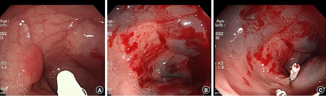
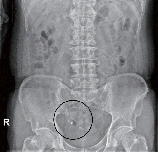
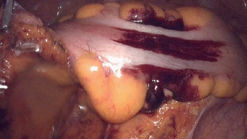
Fig. 4.
Differences in hemoglobin (Hb) changes over time between the control and blood tattooing groups.
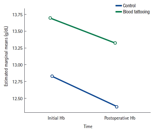
Fig. 5.
Differences in white blood cell count (WBC) changes over time between the control and blood tattooing groups.

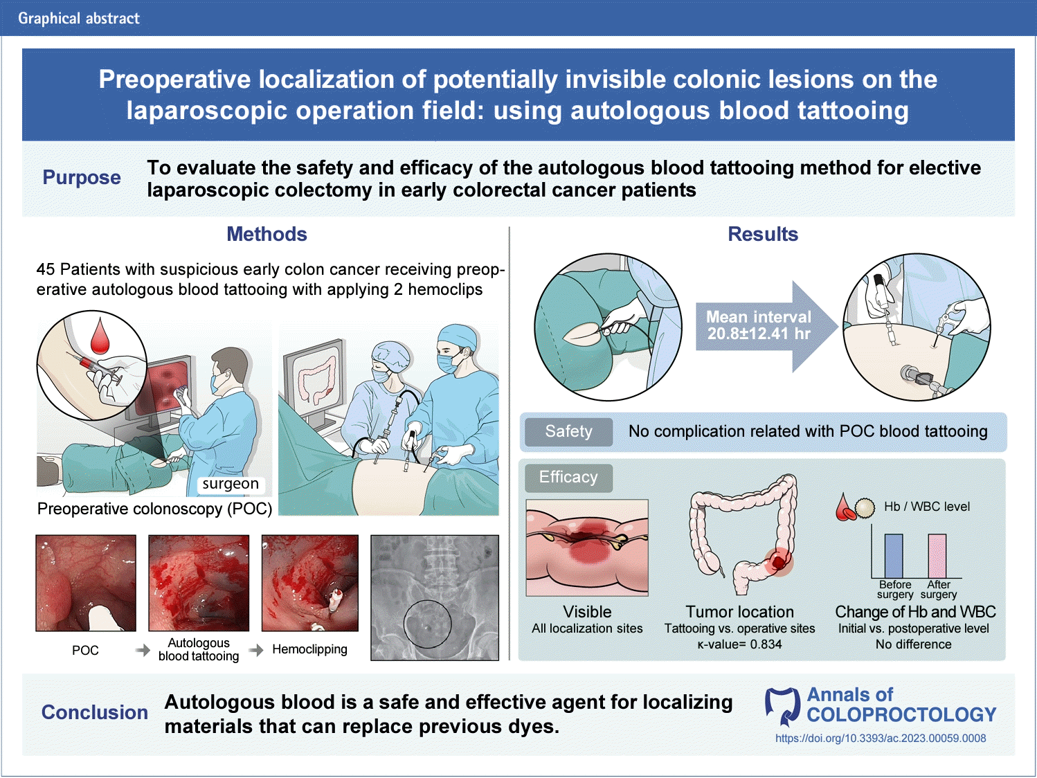
Table 1.
Baseline characteristics (n=45)
Table 2.
The agreement between the colonoscopic (tattooing) and intraoperative findings for colonic lesions requiring localization
Table 3.
The agreement between the initial colonoscopic and intraoperative findings for colonic lesions requiring localization
- References
REFERENCES
1. National Comprehensive Cancer Network (NCCN). NCCN guidelines: colon cancer [Internet]. Version 1.2022. NCCN; 2022 [cited 2022 Jun 11]. Available from: https://www.nccn.org2. Clinical Outcomes of Surgical Therapy Study Group, Nelson H, Sargent DJ, Wieand HS, Fleshman J, Anvari M, et al. A comparison of laparoscopically assisted and open colectomy for colon cancer. N Engl J Med 2004;350:2050–9.
[Article] [PubMed]3. Wang CL, Qu G, Xu HW. The short- and long-term outcomes of laparoscopic versus open surgery for colorectal cancer: a metaanalysis. Int J Colorectal Dis 2014;29:309–20.
[Article] [PubMed]4. Fleshman J, Branda ME, Sargent DJ, Boller AM, George VV, Abbas MA, et al. Disease-free survival and local recurrence for laparoscopic resection compared with open resection of stage II to III rectal cancer: follow-up results of the ACOSOG Z6051 randomized controlled trial. Ann Surg 2019;269:589–95.
[Article] [PubMed] [PMC]5. Cohen JL, Forde KA. Intraoperative colonoscopy. Ann Surg 1988;207:231–3.
[Article] [PubMed] [PMC]6. Letarte F, Webb M, Raval M, Karimuddin A, Brown CJ, Phang PT. Tattooing or not? A review of current practice and outcomes for laparoscopic colonic resection following endoscopy at a tertiary care centre. Can J Surg 2017;60:394–8.
[Article] [PubMed] [PMC]7. Lee SJ, Sohn DK, Han KS, Kim BC, Hong CW, Park SC, et al. Preoperative tattooing using indocyanine green in laparoscopic colorectal surgery. Ann Coloproctol 2018;34:206–11.
[Article] [PubMed] [PMC]8. Aawsaj YM, Kelly S, Slater B. Liver abscess secondary to an endoscopic tattoo in the colon. Ann R Coll Surg Engl 2017;99:e47–8.
[Article] [PubMed] [PMC]9. Jeong O, Cho SB, Joo YE, Ryu SY, Park YK. Novel technique for intraoperative tumor localization during totally laparoscopic distal gastrectomy: endoscopic autologous blood tattooing. Surg Endosc 2012;26:1778–83.
[Article] [PubMed]10. Ponsky JL, King JF. Endoscopic marking of colonic lesions. Gastrointest Endosc 1975;22:42–3.
[Article] [PubMed]11. Park SI, Genta RS, Romeo DP, Weesner RE. Colonic abscess and focal peritonitis secondary to india ink tattooing of the colon. Gastrointest Endosc 1991;37:68–71.
[Article] [PubMed]12. Bahadursingh AM, Driver M, Koenig CL, Longo WE. Inadvertent transmural India ink tattooing simulating intestinal infarction. Am J Surg 2003;185:88–9.
[Article] [PubMed]13. Coman E, Brandt LJ, Brenner S, Frank M, Sablay B, Bennett B. Fat necrosis and inflammatory pseudotumor due to endoscopic tattooing of the colon with india ink. Gastrointest Endosc 1991;37:65–8.
[Article] [PubMed]14. Algoe KK, Chen H, Schned AR, Whiteside JL. Intraperitoneal India ink deposits appearing as endometriosis in a patient with chronic pelvic pain. Obstet Gynecol 2008;112(2 Pt 2):448–50.
[Article]15. Pedersen R, Chen J, Kho KA. Peritoneal and intestinal ink stains from endoscopic tattooing encountered during gynecologic surgery. J Minim Invasive Gynecol 2021;28:1669–70.
[Article] [PubMed]16. Ohdaira T, Konishi F, Nagai H, Kashiwagi H, Shito K, Togashi K, et al. Intraoperative localization of colorectal tumors in the early stages using a marking clip detector system. Dis Colon Rectum 1999;42:1353–5.
[Article] [PubMed]17. Cai Z, Pan R, Ma J, Zheng M. Tumor localization for laparoscopic colorectal resection without endoscopic tattooing. Surg Laparosc Endosc Percutan Tech 2016;26:230–5.
[Article] [PubMed]18. Rockey DC, Paulson E, Niedzwiecki D, Davis W, Bosworth HB, Sanders L, et al. Analysis of air contrast barium enema, computed tomographic colonography, and colonoscopy: prospective comparison. Lancet 2005;365:305–11.
[Article] [PubMed]19. Rosman AS, Korsten MA. Meta-analysis comparing CT colonography, air contrast barium enema, and colonoscopy. Am J Med 2007;120:203–10.
[Article] [PubMed]20. Yeo UD, Sung NS, Roh SJ, Choi WJ, Song KH, Choi IS, et al. The usefulness of preoperative colonoscopy tattooing with autologous blood for localization in laparoscopic colorectal surgery. J Minim Invasive Surg 2020;23:114–9.
[Article] [PubMed] [PMC]21. Yeung JM, Maxwell-Armstrong C, Acheson AG. Colonic tattooing in laparoscopic surgery: making the mark? Colorectal Dis 2009;11:527–30.
[Article] [PubMed]22. Yap R, Ianno D, Burgess A. Colonoscopy localization accuracy for colorectal resections in the laparoscopic era. Am J Surg 2016;212:258–63.
[Article] [PubMed]23. ASGE Technology Committee, Hwang JH, Konda V, Abu Dayyeh BK, Chauhan SS, Enestvedt BK, et al. Endoscopic mucosal resection. Gastrointest Endosc 2015;82:215–26.
[Article] [PubMed]24. Hanson ME, Pickhardt PJ, Kim DH, Pfau PR. Anatomic factors predictive of incomplete colonoscopy based on findings at CT colonography. AJR Am J Roentgenol 2007;189:774–9.
[Article] [PubMed]25. Lee SH, Park YK, Lee DJ, Kim KM. Colonoscopy procedural skills and training for new beginners. World J Gastroenterol 2014;20:16984–95.
[Article] [PubMed] [PMC]26. Manigrasso M, Milone M, Musella M, Venetucci P, Maione F, Elmore U, et al. Preoperative Localization in Colonic Surgery (PLoCoS Study): a multicentric experience on behalf of the Italian Society of Colorectal Surgery (SICCR). Updates Surg 2022;74:137–44.
[Article] [PubMed] [PMC]27. Wexner SD, Cohen SM, Ulrich A, Reissman P. Laparoscopic colorectal surgery: are we being honest with our patients? Dis Colon Rectum 1995;38:723–7.
[Article] [PubMed]28. Ghahremanlou R, Bulut O, Jess P. Localization of colonic tumor in laparoscopic surgery. Intraoperative colonoscopy or preoperative tattoo. Ugeskr Laeger 2005;167:2886–9.
[PubMed]29. Kalady MF. Surgeon involvement in preoperative colorectal tumor localization. World J Surg 2010;34:1592–3.
[Article] [PubMed]30. Azin A, Saleh F, Cleghorn M, Yuen A, Jackson T, Okrainec A, et al. A comparison of endoscopic localization error rate between operating surgeons and referring endoscopists in colorectal cancer. Surg Endosc 2017;31:1318–26.
[Article] [PubMed]31. Leyden JE, Doherty GA, Hanley A, McNamara DA, Shields C, Leader M, et al. Quality of colonoscopy performance among gastroenterology and surgical trainees: a need for common training standards for all trainees? Endoscopy 2011;43:935–40.
[Article] [PubMed]32. Singh S, Singh PP, Murad MH, Singh H, Samadder NJ. Prevalence, risk factors, and outcomes of interval colorectal cancers: a systematic review and meta-analysis. Am J Gastroenterol 2014;109:1375–89.
[Article] [PubMed]