Late Presentation of Anal Canal Duplication in Adults: A Series of Four Rare Cases
Article information
Abstract
Anal canal duplication (ACD) is a very rare condition, especially in adults. Four cases in adults are reported. In three cases, the orifice of duplication was located behind the native anus, and in one case, it was located anteriorly. In all cases, no communication between the anal canal and the tract of duplication was noted. Complete removals of the duplications were done through a perineal approach. Histology showed fibro-muscular tissue lined with a squamous epithelium. The postoperative courses were uneventful.
INTRODUCTION
Anal canal duplication (ACD) is a rare condition that is not easy to recognize [1]. It presents as a perineal orifice that ends with a tract as long as the anal canal and without any communication with the anorectum [2]. ACDs are usually detected early in the life, and only a few cases remain undiagnosed until adulthood. Most of the conditions are reported in children and infants [1]. We report on a series of four cases of ACD in adults.
CASE REPORTS
Case 1
A 20-year-old woman was admitted with a perianal abscess twice, two years and one month earlier. The patient was normal at birth, but developed a painful and warm swelling in postanal area two years before. The abscess was incised and drained. On the second admission, on month earlier, she presented with a perineal abscess at the same region. Incisional drainage of the abscess resulted in a nonhealing perianal fistula. The anus was normal, and a fistula opening was situated about 1 cm posterior to the anal verge. Purulent fluid was being discharged from the fistula. Digital rectal examination revealed a well-defined tubular structure of about 6 cm × 4 cm in size in the retrorectal area. Compression of this mass resulted in pus discharge from the perianal fistula. Magnetic resonance imaging of the perineum showed a tract as long as the anal canal. After the infection had been controlled, the cyst was excised through a perineal approach. There was no connection between the cyst and the anal canal. The tract in the postanal space was dissected off the postanal space, and the cyst, which extended to S2-S3, was excised from all aspects. A hemovac drain was inserted, and the muscles were approximated with absorbable sutures. The postoperative recovery was without complication. Inflamed fibro-muscular tissue lined by a striated squamous epithelium was demonstrated on pathology (Fig. 1).
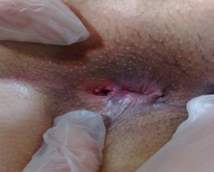
Anal canal duplication in a 20-year-old woman.
Case 2
A 50-year-old woman was admitted to our department for a perineal mass. She had undergone surgery for this mass twice before. Contrast was injected through an opening in the perineum posterior to the anus. It showed a tract that ended with a cyst-like lesion. No connection between the cyst and the canal existed. The whole tract was excised. Histology showed a fibro-muscular tissue lined by a squamous epithelium.
Case 3
A 35-year-old woman presented with a perineal opening at the 12 o'clock position. She had previously undergone surgery for a perineal mass by a gynecologist. After the surgery, an anterior perineal fistula remained. She had a normal anus. Anal function and sphincter tone were normal. Discharge from the opening was noted. She had no history of perianal abscess. Carful inspection of the opening and the tract revealed an anal canal pattern, including a dentate line. Excision of the tract by using cautery dissection around the fistula tract was carried out proximally for a distance of 4 cm. No connection between the tract and the vagina or the rectum was observed. Postoperative recovery was uneventful. Histological examination showed an anal mucosa with congestion and a mild, chronic, nonspecific inflammation.
Case 4
A 24-year-old woman was admitted with a perineal orifice at the 5 o'clock position. This small opening had been noted since birth, but ignored. She had an anus with normal tone in the normal position. She had no history of abscess or discharge from the orifice. Fistulography showed a 4-cm canal with no communication to the rectum. Also, in this case, the tract had an anal canal pattern with a dentate line. Excision of the canal was performed. Histology revealed an anal mucosa. A small hematoma, which was incised and drained, was present at the site of excision (Fig. 2).
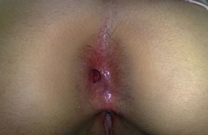
Anal canal duplication in a 24-year-old woman.
DISCUSSION
ACD is a rare developmental lesion. It is a midline lesion that is usually situated behind the native anus; Jacquier et al. [3] defined ACD as "duplications along the posterior side of the anal canal, with a perineal orifice situated just behind the anus at 6 o'clock, with a physical aspect of a duplicate anus." However, this definition does not include ACDs in other positions, as seen in one patient in our report. The case review by Carpentier et al. [1] involved four patients with ACDs in other positions. Therefore, pathologic definition by Ochiai et al. [4] seems to be more appropriate: They defined ACD based on three pathological characteristics: a squamous epithelium in the caudal end, a transitional epithelium in the cranial end, and smooth-muscle cells in the wall of the ACD. ACD is usually asymptomatic [5] and may be missed until adulthood. Some patients present with complications, such as abscesses, recurrent fistulae, and malignant changes later in life [1, 6].
In our report, three patients presented with an abscess and a recurrent fistula, and one patient was asymptomatic. Early diagnosis can be made by simple perineal inspection, which reveals a small opening located in the midline and is usually just behind the normal anus. In our report, a careful inspection in at least two cases, revealed an anal canal-like tract with a dentate line. ACD should be differentiated from rectal duplication or fistula-in-ano. Rectal duplication is situated in the front of or on the rear side of the anorectum, frequently creating a cystic mass [7]. However, the main difference between these two pathologies and an ACD is related to their histologic examination after excision. Imaging assessment confirms the size, type, possible anorectal communication, and mass effect of the ACD.
Removal of the ACD is recommended because of the possibility of infection and malignant change, as reported by Dukes and Glavin [6] in eight of ten adult cases. In our experience, we preferred complete removal of the duplication by using a perineal approach. Lisi et al. [7] also removed the whole lesion completely, but Tiryaki et al. [5], in two cases of ACD, considered simple mucosal stripping with primary repair sufficient to remove the tissue involved in possible future malignancy.
In our research, the diagnosis of ACD was confirmed with the pathologic examination: We found squamous cells and smooth muscle cells in the wall of the ACD. No transitional or columnar cells that might be related to the lengths of the lesions were found in our specimens. Choi and Park [8] suggested that the histological characteristics of an ACD might vary based on the length of the structure. For example, if the depth of the pit was short, one might not see a transitional epithelium at the cranial end.
In conclusion, although ACD is a very rare condition, we must consider it when we encounter a perineal orifice. At diagnosis, surgical removal of the ACD is warranted due to inflammatory complications and late malignant changes.
ACKNOWLEDGMENTS
We would like to express our sincere gratitude to the Tehran University of Medical Sciences.
Notes
No potential conflict of interest relevant to this article was reported.