Warning: fopen(/home/virtual/colon/journal/upload/ip_log/ip_log_2025-12.txt): failed to open stream: Permission denied in /home/virtual/lib/view_data.php on line 95 Warning: fwrite() expects parameter 1 to be resource, boolean given in /home/virtual/lib/view_data.php on line 96 Primary Multiple Mesenteric Liposarcoma of the Transverse Mesocolon
Skip Navigation
Skip to contents

Ann Coloproctol : Annals of Coloproctology

OPEN ACCESS
SEARCH
Search

Articles

Page Path
HOME > Ann Coloproctol > Volume 29(3); 2013 > Article
Case Report
Primary Multiple Mesenteric Liposarcoma of the Transverse Mesocolon
Sandeep Sachidananda, Arunkumar Krishnan1, Raja Ramesh, Sivaprakasam Kuppurao
Annals of Coloproctology 2013;29(3):123-125.
DOI: https://doi.org/10.3393/ac.2013.29.3.123
Published online: June 30, 2013

Department of General Surgery, Stanley Medical College and Hospital, Chennai, India.

1Department of Gastroenterology and Hepatology, Stanley Medical College and Hospital, Chennai, India.

Correspondence to: Arunkumar Krishnan, M.D. Department of Gastroenterology and Hepatology, Stanley Medical College and Hospital, C-137, Karthikeyan Salai, Periyar Nagar, Perambur, Chennai 600082, Tamilnadu State, India. Tel: +91-44-25289595, Fax: +91-442-5289595, dr.arunkumarpillai@gmail.com
• Received: July 17, 2012   • Accepted: October 3, 2012

© 2013 The Korean Society of Coloproctology

This is an open-access article distributed under the terms of the Creative Commons Attribution Non-Commercial License (http://creativecommons.org/licenses/by-nc/3.0/) which permits unrestricted non-commercial use, distribution, and reproduction in any medium, provided the original work is properly cited.

prev next
  • 4,800 Views
  • 28 Download
  • 8 Crossref
  • 8 Scopus
  • Liposarcomas are common tumors arising in the retroperitoneum. However, a primary mesenteric liposarcoma is a rare entity and less than 50 cases have been reported so far. Further, a liposarcoma arising in the transverse mesocolon is very unusual, and cases of multiple primary tumors arising from the transverse mesocolon are extremely rare. We want to report a case of a multiple primary mesenteric liposarcoma arising from the transverse mesocolon in a 63-year-old female who was successfully treated by surgery. Because a primary mesenteric liposarcoma is a rare entity, it should be considered with the differential diagnosis of an abdominal mesenchymal tumor. It can be diagnosed preoperatively by using contrast enhanced computed tomography and magnetic resonance imaging. The treatment for such a liposarcoma is surgical resection with sufficient surgical margin; the role of adjuvant therapy has yet to be defined.
Liposarcomas are the second most common type of soft tissue sarcomas [1]. They are commonly known to occur in the retroperitoneum. A primary mesenteric origin of a liposarcoma is an unusual occurrence and has been reported to occur from the small bowel mesentery. A primary origin from the transverse mesocolon is a rare occurrence [2, 3]. The multiplicity of these tumors arising from the transverse mesocolon in our case makes it a unique one.
A 63-year-old female with no comorbidities presented with gradually progressive, painless, upper abdominal distension and loss of appetite for one year. Physical examination revealed a distended abdomen and epigastrium, as well as right and left hypochondrium fullness extending to the umbilicus. A nontender mass of about 15 cm × 12 cm in size was palpable in the epigastrium, the right and the left hypochondrium, and the umbilical regions. The mass was firm, had a smooth surface, moved from side to side, and fell forward when the patient was in the knee-elbow position. The findings of abdominopelvic ultrasound showed a 12.5 cm × 9.6 cm uniformly echogenic lesion with unclear margins in the midline; the lesion occupied the epigastric and umbilical regions and pushed bowel loops to the periphery.
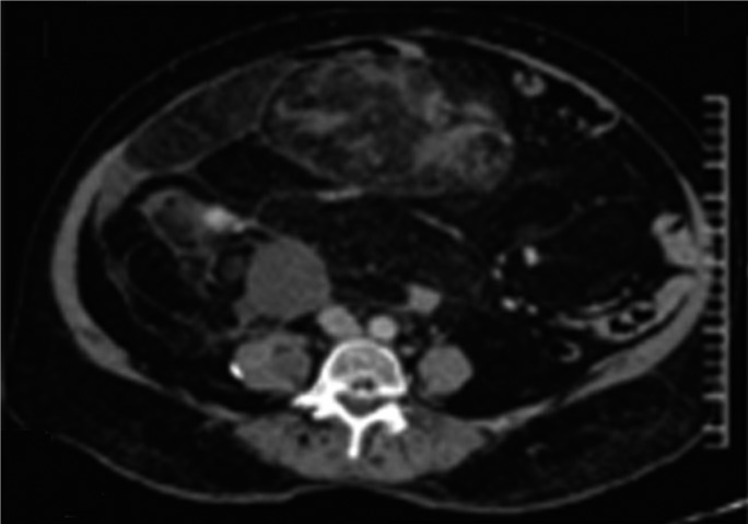
Contrast enhanced computed tomography (CECT) of the abdomen and the pelvis showed a space-occupying lesion in the epigastric/umbilical region measuring 16 cm × 16 cm × 10 cm. The lesion was a nonhomogenous mass with irregular enhancement and displaced the bowel loops (straddling sign, Fig. 1). Bowel loops were located between the tumor and the anterior abdominal wall, suggesting a probable mesenteric origin [4]. No evidence of a retroperitoneal lymphadenopathy was found, and the lesion was reported as a large teratoma occupying the anterior epigastric/umbilical regions with a possible mesenteric origin.
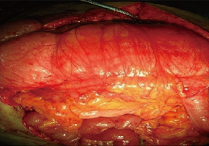
The patient underwent surgery. A midline laparotomy was done, and the peritoneal cavity was explored. A huge lipomatous mass measuring 23 cm × 16 cm × 12 cm was found in the lesser sac over the transverse mesocolon. The lesser sac was opened, and the mass was found to be well defined without any major vascular invasion. The tumor was excised in toto with macroscopically negative margins. On further exploration, multiple such separated masses with sizes ranging from 4 to 7 cm were found to be attached to the transverse mesocolon (Figs. 2 and 3), and each was excised with clear margins. Postoperative period was uneventful. Histopathology showed similar features suggestive of an atypical lipoma or a well-differentiated liposarcoma in all of the excised lesions, suggesting possible multicentricity or the presence of satellite nodules.
The liposarcoma is the second most common soft tissue sarcoma and frequently occurs in the lower extremities and the retroperitoneum. Primary tumors of the mesentery are uncommon. Less than 50 such cases of a primary mesenteric liposarcoma have been reported to date, and very few cases of a liposarcoma of the transverse mesocolon have been reported [2, 3]. A mesenteric liposarcoma is a tumor seen in adults aged 50 to 70 years and is slightly more common in males [5]. It is usually nonmetastasizing and locally aggressive, presents as a deep-seated mass located more towards the root of the mesentery rather than near the intestine, and can grow to a large size. It is often painless and presents with non-specific vague symptoms like anorexia, early satiety, and abdominal distention, and occasionally can cause subacute intestinal obstruction or volvulus. The main differential diagnosis for mesenteric tumors is a malignant lymphoma, leiomyosarcoma, or pelvic lipomatosis/lipodystrophy [5].
A liposarcoma is histologically defined as a tumor composed of lipoblasts and can secrete substances such as granulocyte-colony-stimulating factor (G-CSF) [3]. For diagnosis, computed tomographic (CT) scans and magnetic resonance imaging (MRI) can provide information regarding the important structures, as well as the tissue characteristics, of the tumor. The characteristics of a liposarcoma on CT images are nonhomogeneity, infiltration or poor marginalization, and higher CT contrast enhancement than normal fats. The enhancement on CT changes according to the histological grade [1]. In the present case, CT scans revealed a tumor with nonhomogeneous, irregular enhancement. The treatment strategy for a mesenteric liposarcoma is surgical resection with sufficient surgical margin. Complete surgical resection at the time of primary presentation is likely to improve the chance for long-term survival, as well as distant recurrence-free survival, and positive surgical margins are the main predictors of local relapse [5]. The roles of preoperative and postoperative adjuvant chemotherapy and radiotherapy are not very clear. Although reports advocate their usefulness, their survival benefits in the case of a liposarcoma has yet to be established. The key chemotherapy drug is doxorubicin while Ifosfamide, dacarbazine and cisplatin have also been used [4].
In conclusion, although a primary mesenteric liposarcoma is a rare entity, it should be considered in the differential diagnosis of abdominal mesenchymal tumors. It usually presents as a slow-growing tumor that can attain large sizes and cause intestinal obstruction. It can be diagnosed preoperatively by using CECT and MRI. The treatment of choice for such a liposarcoma is surgical resection with sufficient surgical margin, but the role of adjuvant therapy has yet to be defined.

No potential conflict of interest relevant to this article was reported.

  • 1. Hirakoba M, Kume K, Yamasaki M, Kanda K, Yoshikawa I, Otsuki M. Primary mesenteric liposarcoma successfully diagnosed by preoperative imaging studies. Intern Med 2007;46:373–375. ArticlePubMed
  • 2. Yoshiyuki M, Kazuhiko N, Tomoo O, Yoshihiro N, Fujio W, Takeshi K. A case of primary liposarcoma arisen from the mesentery of the transverse colon. J Jpn Surg Assoc 2003;64:2221–2224.Article
  • 3. Nakamura A, Tanaka S, Takayama H, Sakamoto M, Ishii H, Kusano M, et al. A mesenteric liposarcoma with production of granulocyte colony-stimulating factor. Intern Med 1998;37:884–890. ArticlePubMed
  • 4. Ishiguro S, Yamamoto S, Chuman H, Moriya Y. A case of resected huge ileocolonic mesenteric liposarcoma which responded to pre-operative chemotherapy using doxorubicin, cisplatin and ifosfamide. Jpn J Clin Oncol 2006;36:735–738. ArticlePubMed
  • 5. Burgohain J, Kathiresan N, Satheesan B. Dumbbell-shaped mesenteric liposarcoma: a case report with review of the literature. Internet J Surg 2008 cited 2013 Jun 1. 15(2): [about 2 p.]. Available from: http://archive.ispub.com/journal/the-internet-journal-of-surgery/volume-15-number-2/dumbbell-shaped-mesenteric-liposarcoma-a-case-report-with-review-of-the-literature.html#sthash.3W4jux8m.dpbs
Fig. 1
Contrast enhanced computed tomography of the abdomen showing a large tumor with nonhomogenous, irregular enhancement and the presence of a bowel loop between the tumor and the anterior abdominal wall (straddling sign).
ac-29-123-g001.jpg
Fig. 2
Multiple lipomatous tumors arising from transverse mesocolon.
ac-29-123-g002.jpg
Fig. 3
Intraoperative picture showing a large tumor arising from the transverse mesocolon.
ac-29-123-g003.jpg

Figure & Data

References

    Citations

    Citations to this article as recorded by  
    • Giant transverse colonic mesenteric mucinous liposarcoma combined with rectal cancer and aortic coarctation: A case report and review of literature
      Meng Wang, Jian Sun, Zhi-Qiang Song, Xi-Qi Chen, Guang-Dong Xie, Yong Zhu, Yong-Kun Zhou
      World Journal of Gastrointestinal Surgery.2025;[Epub]     CrossRef
    • Clinicopathological Characteristics and Survival Analysis of Primary Mesenteric Liposarcoma: A Retrospective Study
      Zeki Ogut, Adem Tuncer, Yasin Dalda, Harika Gozde Gozukara Bag, Mehmet Bugra Bozan
      Medicina.2025; 61(11): 1984.     CrossRef
    • Dedifferentiated liposarcoma of small bowel mesentery: a rare case report
      Sunil Dhakal, Ijendra Prajapati
      Journal of Surgical Case Reports.2022;[Epub]     CrossRef
    • Giant mesenteric myxoid liposarcoma: Challenges of diagnosis and treatment
      Rania Mokfi, Farah Boutaggount, Meryem Maskrout, Ghizlane Rais
      Radiology Case Reports.2022; 17(11): 4227.     CrossRef
    • Dedifferentiated liposarcoma in the abdominal cavity: a case report
      Marina Kuroda, Reiko Yamada, Takamitsu Tanaka, Junya Tsuboi, Misaki Nakamura, Masaki Katsurahara, Yasuhiko Hamada, Kyosuke Tanaka, Noriyuki Horiki, Hayato Nakagawa
      Clinical Journal of Gastroenterology.2022; 15(6): 1094.     CrossRef
    • The colorectal surgeon's liposarcoma
      Alfred Wong, Han‐Kuang Chen, Michael Levitt
      ANZ Journal of Surgery.2018;[Epub]     CrossRef
    • A mesenteric solid tumor with unusual features in a young male: A case report
      Yoshitoshi Ichikawa, Tamaki Maeda, Gaku Mizojiri, Satoshi Ishikawa, Takaomi Hagi, Kimiaki Hattori, Hiroshi Oka
      Molecular and Clinical Oncology.2017; 7(3): 355.     CrossRef
    • Multiple mesenteric well-differentiated liposarcoma complicated by purulent inflammation: A case report
      WEI GAO, HUAIZHOU WANG, JINYU LIU, FUJIANG WANG, JIANJUN DONG, JUNZU GENG
      Oncology Letters.2015; 9(3): 1333.     CrossRef

    • Cite this Article
      Cite this Article
      export Copy Download
      Close
      Download Citation
      Download a citation file in RIS format that can be imported by all major citation management software, including EndNote, ProCite, RefWorks, and Reference Manager.

      Format:
      • RIS — For EndNote, ProCite, RefWorks, and most other reference management software
      • BibTeX — For JabRef, BibDesk, and other BibTeX-specific software
      Include:
      • Citation for the content below
      Primary Multiple Mesenteric Liposarcoma of the Transverse Mesocolon
      Ann Coloproctol. 2013;29(3):123-125.   Published online June 30, 2013
      Close
    • XML DownloadXML Download
    Figure
    • 0
    • 1
    • 2
    Primary Multiple Mesenteric Liposarcoma of the Transverse Mesocolon
    Image Image Image
    Fig. 1 Contrast enhanced computed tomography of the abdomen showing a large tumor with nonhomogenous, irregular enhancement and the presence of a bowel loop between the tumor and the anterior abdominal wall (straddling sign).
    Fig. 2 Multiple lipomatous tumors arising from transverse mesocolon.
    Fig. 3 Intraoperative picture showing a large tumor arising from the transverse mesocolon.
    Primary Multiple Mesenteric Liposarcoma of the Transverse Mesocolon

    Ann Coloproctol : Annals of Coloproctology Twitter Facebook
    TOP