Warning: fopen(/home/virtual/colon/journal/upload/ip_log/ip_log_2025-11.txt): failed to open stream: Permission denied in /home/virtual/lib/view_data.php on line 95 Warning: fwrite() expects parameter 1 to be resource, boolean given in /home/virtual/lib/view_data.php on line 96 Giant Ascending Colonic Diverticulum Presenting With Intussusception
Skip Navigation
Skip to contents

Ann Coloproctol : Annals of Coloproctology

OPEN ACCESS
SEARCH
Search

Articles

Page Path
HOME > Ann Coloproctol > Volume 29(5); 2013 > Article
Case Report
Giant Ascending Colonic Diverticulum Presenting With Intussusception
Ho Jin Kim, Jin Ha Kim, Ok In Moon, Kyung Jong Kim
Annals of Coloproctology 2013;29(5):209-212.
DOI: https://doi.org/10.3393/ac.2013.29.5.209
Published online: October 31, 2013

Department of Surgery, Chosun University School of Medicine, Gwangju, Korea.

Correspondence to: Kyung Jong Kim, M.D. Department of Surgery, Chosun University Medical School, 309 Pilmun-daero, Dong-gu, Gwangju 501-759, Korea. Tel: +82-62-220-3983, Fax: +82-62-228-3441, kjkim@chosun.ac.kr
• Received: May 24, 2013   • Accepted: July 30, 2013

© 2013 The Korean Society of Coloproctology

This is an open-access article distributed under the terms of the Creative Commons Attribution Non-Commercial License (http://creativecommons.org/licenses/by-nc/3.0/) which permits unrestricted non-commercial use, distribution, and reproduction in any medium, provided the original work is properly cited.

prev next
  • 5,101 Views
  • 54 Download
  • 10 Crossref
  • 11 Scopus
  • Diverticular disease of the colon is a common disease, and its incidence is increasing gradually. A giant colonic diverticulum (GCD) is a rare entity and is defined as a diverticulum greater than 4 cm in size. It mainly arises from the sigmoid colon, and possible etiology is a ball-valve mechanism permitting progressive enlargement. A plain abdominal X-ray can be helpful to make a diagnosis initially, and a barium enema and abdominal computed tomography may confirm the diagnosis. Surgical intervention is a definite treatment for a GCD. We report a case of an ascending GCD presenting with intussusception in a young adult.
Diverticular disease is a common disorder occurring in elderly people, and its incidence is increasing. No more than 20% of the patients with colonic diverticulum have symptoms, and of those who have symptoms, 25% experience complications such as abscess, perforation, stricture or fistula. Adult intussusception represents 5% of all cases of intussusception and accounts for only 1-5% of intestinal obstructions in adults [1]. Almost 90% of intussusceptions in adult are secondary to a carcinoma, polyp, stricture or benign neoplasm, including a colonic diverticulum [2]. A giant colonic diverticulum (GCD) is a rare entity of disease and is defined as a diverticulum greater than 4 cm in size [3]. More than 90% are antimesenteric and arise from the sigmoid colon [4]. Several GCDs have been reported, almost all of which developed at old age and in the sigmoid colon [5, 6]. We report a case of a GCD, which was removed by using a laparoscopic colectomy. To our knowledge, this is the first case of an ascending GCD in a young adult that suggests a congenital origin in pathogenesis and presented with intussusceptions.
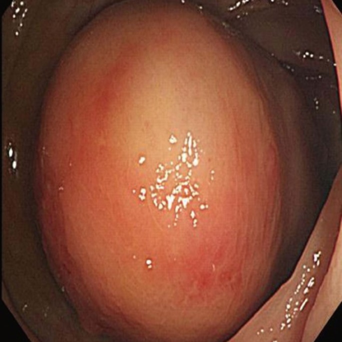
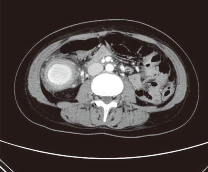
A 30-year-old female was transferred to Chosun University Hospital with a 1-month history of left lower quadrant pain. The laboratory examination at admission was unremarkable. Physical examination revealed left lower quadrant tenderness with a palpable abdominal mass. Plain abdomen X-ray presented a gas-filled cavity in the middle right abdomen. The computed tomography scan from an outside hospital showed a target sign suggesting intussusception in the mid to lower colon on the left side (Fig. 1). The intussusception was soon resolved spontaneously without any procedure. To evaluate the lesion, we performed a colonoscopic examination, which revealed a huge submucosal tumor (SMT)-like lesion in the ascending colon (Fig. 2). The abdominal computed tomographic scan was checked again for further assessment of the SMT-like lesion and showed 4.2-cm-sized encapsulated fluid of high density with a thick wall on the right side of abdomen (Fig. 3).
A laparoscopic right hemicolectomy was performed because of uncertainty of the diagnosis and for a one-stage treatment. On the operation findings, the lesion which was 5 cm × 5 cm in size was located in the mid portion of the ascending colon and extended to the hepatic flexure. Also, multiple enlarged lymph nodes were found around the ileocolic and the middle colic vessel and were removed by using an en bloc resection. Gross finding of the removed specimens showed a large diverticulum on the ascending colon (Fig. 4A). Histologic examination of the diverticulum showed the full thickness of the bowel wall from the mucosa to the serosa (Fig. 4B). The patient made a quick recovery and was discharged 7 days after surgery without any complication.
Colonic diverticular disease is a common condition and rarely presents as a singular giant diverticulum. A GCD, which is greater than 4 cm in size, is an uncommon manifestation of colonic diverticular disease and mainly occurs solitarily in the sigmoid colon [5, 7]. The pathogenesis of a GCD is not well established. Possible theory supports a ball-valve mechanism through a tiny communicating neck, which causes air entrapment so that the diverticulum increases in size. Another theory postulates that the formation and the enlargement of a GCD is secondary to gas-forming organisms [8]. There are different types of GCDs. Steenvoorde et al. [9] described a histological classification of three distinct subtypes of GCDs. Type I is a pseudo-diverticulum containing remnants of the muscularis mucosa and the muscularis propria, and this type of diverticulum is found in colonic diverticular disease. Type II is an inflammatory GCD resulting from a local perforation of the mucosa with a persistent abscess cavity retaining contact with the colonic lumen, and the type III is a true diverticulum containing all layers of a normal bowel.
The clinical presentation of a GCD can be asymptomatic, but patients present most commonly with abdominal pain, constipation, or a palpable, nontender, abdominal mass [9, 10]. A plain abdominal X-ray is used as the first line for diagnosis of a GCD. It is the simplest and most readily available tool and can reveal a large air-filled structure with or without fluid levels. A barium enema can demonstrate communication with the large bowel lumen in the majority of cases [11], but fails in approximately one-third of reported cases [7, 9]. The differential diagnosis for a GCD includes sigmoid or caecal volvulus, a duplication cyst or giant Meckel diverticulum, and pneumatosis cystoides intestinalis [3].
A plain supine abdominal X-ray is the simplest and most readily available diagnostic tool and should be used as the first line in suspected cases because it can reveal a large air-filled structure (balloon-like appearance) with or without fluid levels. Computed tomography shows a smooth, thick-walled, air-filled structure intimately associated with the colon [8]. Colonoscopic evaluation is difficult in diagnosing a GCD. The ostium between the diverticulum and the colon is frequently too small to be detected [7-9]. GCD cases that have spontaneously resolved with conservative treatment have been reported; nevertheless, the gold standard of management of a GCD is a surgical resection and a primary anastomosis to exclude an underlying malignancy because adenocarcinomas arising from GCDs have been reported previously [9].
In the era of minimal invasive surgery, a laparoscopic colonic resection including the lesion and a primary anastomosis may be feasible in most patients. Laparoscopic surgery has advantages of fast recovery, reduced hospital stay, less pain and better cosmesis [11, 12]. Our case has some differences compared with previous cases. The lesion in our patient occurred in the ascending colon, not the sigmoid colon even though. The histological classification of our case was a true diverticulum, and the origin of diverticulum in our case might have been congenital because the patient was a young adult, unlike most GCD cases where the patients are much older. Our patient was treated with a laparoscopic right hemicolectomy, and there have been no symptoms or signs of recurrence since then (2 years).

No potential conflict of interest relevant to this article was reported.

  • 1. Marinis A, Yiallourou A, Samanides L, Dafnios N, Anastasopoulos G, Vassiliou I, et al. Intussusception of the bowel in adults: a review. World J Gastroenterol 2009;15:407–411. ArticlePubMedPMC
  • 2. Weilbaecher D, Bolin JA, Hearn D, Ogden W 2nd. Intussusception in adults: review of 160 cases. Am J Surg 1971;121:531–535. ArticlePubMed
  • 3. Beddy D, DeBlacam C, Mehigan B. An unusual cause of an acute abdomen: a giant colonic diverticulum. J Gastrointest Surg 2010;14:2016–2017. ArticlePubMed
  • 4. Custer TJ, Blevins DV, Vara TM. Giant colonic diverticulum: a rare manifestation of a common disease. J Gastrointest Surg 1999;3:543–548. ArticlePubMed
  • 5. Kang JY, Hoare J, Tinto A, Subramanian S, Ellis C, Majeed A, et al. Diverticular disease of the colon--on the rise: a study of hospital admissions in England between 1989/1990 and 1999/2000. Aliment Pharmacol Ther 2003;17:1189–1195. ArticlePubMed
  • 6. Kang JY, Melville D, Maxwell JD. Epidemiology and management of diverticular disease of the colon. Drugs Aging 2004;21:211–228. ArticlePubMed
  • 7. Levi DM, Levi JU, Rogers AI, Bergau DK, Wenger J. Giant colonic diverticulum: an unusual manifestation of a common disease. Am J Gastroenterol 1993;88:139–142. PubMed
  • 8. de Oliveira NC, Welch JP. Giant diverticula of the colon: a clinical assessment. Am J Gastroenterol 1997;92:1092–1096. PubMed
  • 9. Steenvoorde P, Vogelaar FJ, Oskam J, Tollenaar RA. Giant colonic diverticula: review of diagnostic and therapeutic options. Dig Surg 2004;21:1–6. ArticlePubMed
  • 10. Scott DA, Glancy S. Spontaneous resolution of a giant colonic diverticulum. Clin Radiol 2008;63:833–835. ArticlePubMed
  • 11. Choong CK, Frizelle FA. Giant colonic diverticulum: report of four cases and review of the literature. Dis Colon Rectum 1998;41:1178–1185. ArticlePubMed
  • 12. Galandiuk S, Fazio VW. Pneumatosis cystoides intestinalis: a review of the literature. Dis Colon Rectum 1986;29:358–363. ArticlePubMed
Fig. 1
Initial abdominal computed tomographic scan from an outside hospital showing the target sign in the left mid to lower abdomen.
ac-29-209-g001.jpg
Fig. 2
Colonoscopic finding showing a large, smooth-surfaced, round mass lesion, mimicking a submucosal tumor, near the hepatic flexure.
ac-29-209-g002.jpg
Fig. 3
Follow-up abdomen computed tomographic (CT) scan. Comparing to the previous CT scan, the large high-density mass had moved to the right side of the abdomen, suggesting an ascending colonic mass.
ac-29-209-g003.jpg
Fig. 4
Pathophysiology. Panel A shows a large diverticulum in the gross specimen. Panel B exhibits the true diverticulum and shows the full thickness of the bowel wall from the mucosa to the serosa in a low-power field (H&E, × 100). The white arrow indicates an outpouching of the diverticulum. Panel C shows amorphous materials, consistent with a fecalith, in the diverticular lumen (H&E, × 100).
ac-29-209-g004.jpg

Figure & Data

References

    Citations

    Citations to this article as recorded by  
    • Intussusception secondary to retroflexion of a proximal jejunal diverticulum, leading to Type 3 vagal indigestion with severe hypochloraemia in an adult Simmental bull (Bos taurus)
      Liam A. Wilson, Rob F. Kelly, Adrian W. Philbey
      Veterinary Record Case Reports.2024;[Epub]     CrossRef
    • A case report of impacted fecalith within mucosal pouch: an unusual cause of colocolic intussusception
      Douglas Greer, Adrian Fernandez
      Annals of Coloproctology.2024; 40(Suppl 1): S15.     CrossRef
    • Popping the Balloon: A Giant Colonic Diverticulum Complicated by Bladder Neck Compression
      M. C. Ripoli, A. Lauro, S. Vaccari, G. Mastrocola, A. Lanci-Lanci, V. D’Andrea, I. R. Marino, M. Cervellera, V. Tonini
      Digestive Diseases and Sciences.2021; 66(1): 41.     CrossRef
    • Laparoscopic resection of a giant colonic diverticulum – the ‘lifting balloon’ sign – a video vignette
      C. Rodríguez‐Otero Luppi, M. Rodríguez Blanco, J. Bollo Rodríguez, A. Méndez, J. Merlo Más
      Colorectal Disease.2019; 21(9): 1096.     CrossRef
    • Intussusception caused by an inverted colonic diverticulum: a case report
      Bei Zhang, Jiping Wang, Xiaoguang Li, Zhuo Wang, Yangjiao Zhang, Hao Yang
      Journal of Medical Case Reports.2018;[Epub]     CrossRef
    • Adult intussusception secondary to diverticular disease
      Habib Syed, Labib Syed, Umesh Parampalli, Mokhtar Uheba
      BMJ Case Reports.2018; 2018: bcr-2018-226678.     CrossRef
    • Laparoskopische Resektion eines Riesenkolondivertikels
      P. J. Roch, T. Friedrich, R. Bönninghoff, D. Dinter, A. Rickert
      Der Chirurg.2017; 88(8): 682.     CrossRef
    • Giant colonic diverticulum: radiographic and MDCT characteristics
      Abdel-Rauf Zeina, Ahmad Mahamid, Alicia Nachtigal, Itamar Ashkenazi, Mika Shapira-Rootman
      Insights into Imaging.2015; 6(6): 659.     CrossRef
    • Giant colonic diverticulum: Clinical presentation, diagnosis and treatment: Systematic review of 166 cases
      Giuseppe Nigri
      World Journal of Gastroenterology.2015; 21(1): 360.     CrossRef
    • Giant Colonic Diverticulum: a Rare Diagnostic and Therapeutic Challenge of Diverticular Disease
      Ryan Macht, Holly K. Sheldon, P. Marco Fisichella
      Journal of Gastrointestinal Surgery.2015; 19(8): 1559.     CrossRef

    • Cite this Article
      Cite this Article
      export Copy Download
      Close
      Download Citation
      Download a citation file in RIS format that can be imported by all major citation management software, including EndNote, ProCite, RefWorks, and Reference Manager.

      Format:
      • RIS — For EndNote, ProCite, RefWorks, and most other reference management software
      • BibTeX — For JabRef, BibDesk, and other BibTeX-specific software
      Include:
      • Citation for the content below
      Giant Ascending Colonic Diverticulum Presenting With Intussusception
      Ann Coloproctol. 2013;29(5):209-212.   Published online October 31, 2013
      Close
    • XML DownloadXML Download
    Figure
    • 0
    • 1
    • 2
    • 3
    Giant Ascending Colonic Diverticulum Presenting With Intussusception
    Image Image Image Image
    Fig. 1 Initial abdominal computed tomographic scan from an outside hospital showing the target sign in the left mid to lower abdomen.
    Fig. 2 Colonoscopic finding showing a large, smooth-surfaced, round mass lesion, mimicking a submucosal tumor, near the hepatic flexure.
    Fig. 3 Follow-up abdomen computed tomographic (CT) scan. Comparing to the previous CT scan, the large high-density mass had moved to the right side of the abdomen, suggesting an ascending colonic mass.
    Fig. 4 Pathophysiology. Panel A shows a large diverticulum in the gross specimen. Panel B exhibits the true diverticulum and shows the full thickness of the bowel wall from the mucosa to the serosa in a low-power field (H&E, × 100). The white arrow indicates an outpouching of the diverticulum. Panel C shows amorphous materials, consistent with a fecalith, in the diverticular lumen (H&E, × 100).
    Giant Ascending Colonic Diverticulum Presenting With Intussusception

    Ann Coloproctol : Annals of Coloproctology Twitter Facebook
    TOP