Warning: fopen(/home/virtual/colon/journal/upload/ip_log/ip_log_2024-11.txt): failed to open stream: Permission denied in /home/virtual/lib/view_data.php on line 95 Warning: fwrite() expects parameter 1 to be resource, boolean given in /home/virtual/lib/view_data.php on line 96 A case report of an unexpected colonic polyp: cavernous hemangioma
Skip Navigation
Skip to contents

Ann Coloproctol : Annals of Coloproctology

OPEN ACCESS
SEARCH
Search

Articles

Page Path
HOME > Ann Coloproctol > Volume 39(3); 2023 > Article
Case Report
Benign bowel disease
A case report of an unexpected colonic polyp: cavernous hemangioma
Miguel Mascarenhas*orcid, Rui Morais*, Regina Teixeira, Guilherme Macedo
Annals of Coloproctology 2023;39(3):280-282.
DOI: https://doi.org/10.3393/ac.2020.00535.0076
Published online: July 13, 2021

Department of Gastroenterology, Centro Hospitalar São João, Porto, Portugal

Correspondence to: Miguel Mascarenhas, MD Department of Gastroenterology, Centro Hospitalar São João, Alameda Prof. Hernâni Monteiro, Porto 4200-319, Portugal Email: miguelmascarenhassaraiva@gmail.com
*Miguel Mascarenhas and Rui Morais contributed equally to this study as co-first authors.
• Received: June 24, 2020   • Revised: September 11, 2020   • Accepted: May 9, 2021

Copyright © 2023 The Korean Society of Coloproctology

This is an Open Access article distributed under the terms of the Creative Commons Attribution Non-Commercial License (http://creativecommons.org/licenses/by-nc/4.0/) which permits unrestricted non-commercial use, distribution, and reproduction in any medium, provided the original work is properly cited.

prev next
  • 3,713 Views
  • 124 Download
  • 2 Web of Science
  • 4 Crossref
  • 3 Scopus
  • Cavernous hemangiomas of the colon are rare, benign vascular lesions, and the site most commonly affected is the rectosigmoid junction. Surgical treatment is recommended for large diffuse lesions but in the presence of pedunculated lesions, endoscopic resection should be preferred if possible. We report a case of a 65-year-old man referred for colonoscopy after positive fecal occult blood, that revealed at the level of the sigmoid colon, a wide base pedunculated polyp (35 mm) occupying more than half of the lumen, with the covering mucosa with a vinous appearance. In order to remove the lesion, a detachable snare was placed and polypectomy was performed. During the procedure, the detachable snare was cut with active bleeding, controlled after clip placement and diluted adrenaline injection. Afterwards, histology revealed a polypoid lesion with a hyperplastic mucosa and submucosal plane expanded by numerous thick-walled vessels in the context of a cavernous colonic hemangioma.
Cavernous hemangiomas of the colon are rare, benign vascular lesions, and the site most commonly affected is the rectosigmoid junction. Surgical treatment is recommended for large diffuse lesions but in the presence of pedunculated lesions, endoscopic resection should be preferred if possible.
We report a case of a 65-year-old man referred for colonoscopy after positive fecal occult blood.
A 65-year-old man with no relevant personal history and no usual medication was referred for colonoscopy after positive fecal occult blood test. He had no symptoms and blood tests revealed no signs of anemia or iron deficiency.
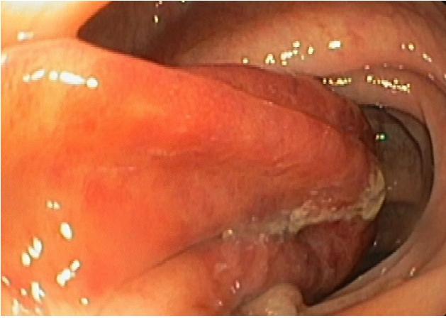
Colonoscopy was performed and revealed, at the level of the sigmoid colon, a wide base pedunculated polyp (35 mm) occupying more than half of the lumen, with the covering mucosa with a vinous appearance (Fig. 1).
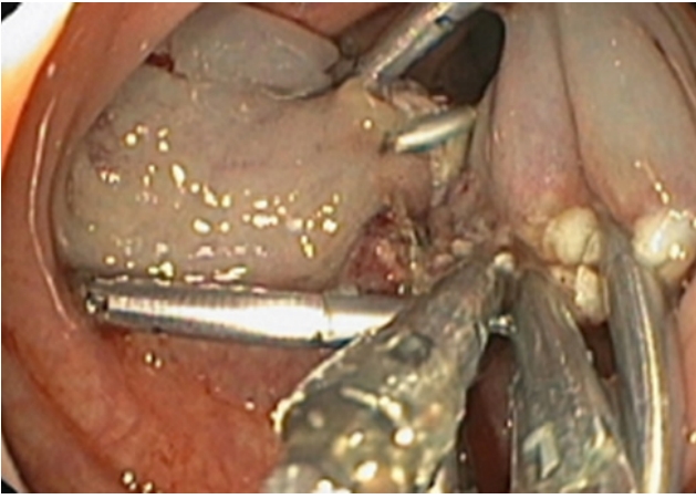
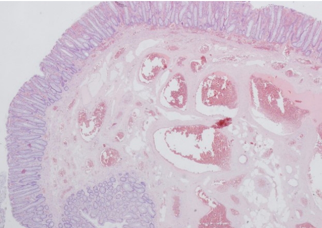
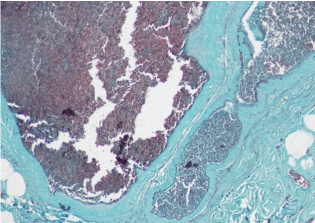
In order to remove the lesion, a detachable snare was placed and hot-snare polypectomy was performed. During the procedure, the detachable sna re was inadvertently cut, with active bleeding that was controlled after clip placement and diluted adrenaline injection (Fig. 2). The patient was admitted for surveillance after the procedure and was discharged after 1 day and did not have bleeding recurrence during the follow-up. The anatomopathological report described a polypoid lesion with hyperplastic mucosa and submucosal plane expanded by numerous thick-walled vessels in the context of cavernous colonic hemangioma (Figs. 3, 4). Colonoscopy was repeated after 3 months and the scar of the previous polypectomy was observed, without evidence of residual lesion.
Ethics statements
Written informed consent was obtained for publication of this case report and accompanying images.
Gastrointestinal hemangiomas are a rare, benign clinical entity that may present as overt or occult bleeding and can be distributed throughout the intestinal digestive system, or present as a singular cavernous hemangioma or malformation [1]. Regarding colonic cavernous hemangiomas the site most commonly affected is the rectosigmoid junction [2].
Colonoscopy is an invaluable tool, being both diagnostic and potentially therapeutic, with cyanoacrylate glue injection, endoscopic mucosal resection, and a variety of thermal modes, including argon plasma coagulation, being part of the therapeutic armamentarium that can possible be used to ablate this type of lesion [3]. Furthermore, there are case reports that validate endoscopic submucosal dissection as an efficient alternative for the complete removal of large colonic cavernous hemangiomas [4]. Surgical treatment is recommended for large diffuse lesions but in the presence of pedunculated lesions, endoscopic resection is a viable and less invasive option [5]. After a PubMed database search, we conclude that this is the second case report of successful removal of pedunculated colonic hemangioma by endoscopic snare polypectomy.
Endoscopic resection does have a significant probability of bleeding and a relevant bowel perforation risk, but it may constitute a less invasive alternative to surgery for smaller and focal vascular lesions such as cavernous hemangioma. For selected cases, we believe that endoscopic resection may have a therapeutic role, before contemplating surgery. Clinical surveillance after the procedure, as well as follow-up endoscopic surveillance, are essential to the safety and success of the procedure.
In the present case, the lesion did not have the typical angiomatous aspect, which may be explained by the presence of hyperplastic mucosal alterations with the presence of expanded vessels at the submucosa level. This case report highlights the importance of a careful endoscopic approach with adequate bleeding prophylaxis for the successful and safe removal of this lesion.

CONFLICT OF INTEREST

No potential conflict of interest relevant to this article was reported.

FUNDING

None.

Fig. 1.
Ileocolonoscopy revealed a wide base pediculated polyp (35 mm) occupying more than half of the lumen, covered by discrete vinous aspect mucosa.
ac-2020-00535-0076f1.jpg
Fig. 2.
An endoloop was placed and an endoscopy polypectomy with diathermic loop was performed. During the procedure, endoloop was cut with active hemorrhage, controlled after clip placement, and diluted adrenaline injection.
ac-2020-00535-0076f2.jpg
Fig. 3.
Polypoid lesion with hyperplastic changes and submucosal plane expanded by numerous thick-walled vessels (hematoxylin-eosin, × 20).
ac-2020-00535-0076f3.jpg
Fig. 4.
Elastic layer enhanced by orcein histochemical staining (× 100).
ac-2020-00535-0076f4.jpg
  • 1. Yoo S. GI-associated hemangiomas and vascular malformations. Clin Colon Rectal Surg 2011;24:193–200.ArticlePubMedPMC
  • 2. Ganesananthan S, Barlow J, Durai D, Hawthorne AB. Multiple venous malformations in the left colon and rectum: a long-standing case managed conservatively and an update of current literature. BMJ Case Rep 2019;12:e227700.ArticlePubMedPMC
  • 3. Leite S, Veloso R, Pinho R, Silva AP, Pinto J, Afonso M, et al. Cavernous hemangiomas limited to the sigmoid colon. Gastrointest Endosc 2010;72:1064–5.ArticlePubMed
  • 4. Chen K, Yan M, Liu F. Successful endoscopic submucosal dissection of a large cavernous hemangioma in the colon. Gastrointest Endosc 2010;72:1064–5.ArticlePubMed
  • 5. Ogasawara N, Suzuki M, Adachi K, Yamaguchi Y, Yamamoto S, Hijikata Y, et al. Endoscopic resection of a pedunculated cavernous hemangioma of the sigmoid colon: a case report. Case Rep Gastroenterol 2019;13:418–22.ArticlePubMedPMC

Figure & Data

References

    Citations

    Citations to this article as recorded by  
    • Endoscopic Resection of a Cavernous Hemangioma in the Sigmoid Colon: A Case Report
      Noora Al-Khater, Mohamed Mohamed, Afra Juma, Faisal Abubaker, Sameer Ansari
      Gastro Hep Advances.2024; 3(3): 396.     CrossRef
    • Anorectal hemangioma, a rare cause of lower gastrointestinal bleeding, treated with selective embolization: A case report
      Barbora Pospisilova, Jaromir Frydrych, Antonin Krajina, Julius Örhalmi, Ivana M Kajzrlikova, Petr Vitek
      World Journal of Gastrointestinal Surgery.2024; 16(8): 2735.     CrossRef
    • Spontaneous colonic perforation of a cavernous hemangioma during colonoscopy
      María Florencia Álvarez, Domingo Cesar Balderramo, Florencia Defanti, Mariano Antonio Higa
      Gastroenterología y Hepatología.2022; 45: 95.     CrossRef
    • A giant hemangioma of the sigmoid colon as a cause of lower gastrointestinal bleeding in a young man
      Lucas Fair, Benjamin Gough, Adatee Oknokwo, Ronney Stadler
      Baylor University Medical Center Proceedings.2022; 35(6): 852.     CrossRef

    • PubReader PubReader
    • ePub LinkePub Link
    • Cite this Article
      Cite this Article
      export Copy Download
      Close
      Download Citation
      Download a citation file in RIS format that can be imported by all major citation management software, including EndNote, ProCite, RefWorks, and Reference Manager.

      Format:
      • RIS — For EndNote, ProCite, RefWorks, and most other reference management software
      • BibTeX — For JabRef, BibDesk, and other BibTeX-specific software
      Include:
      • Citation for the content below
      A case report of an unexpected colonic polyp: cavernous hemangioma
      Ann Coloproctol. 2023;39(3):280-282.   Published online July 13, 2021
      Close
    • XML DownloadXML Download
    Figure
    • 0
    • 1
    • 2
    • 3
    A case report of an unexpected colonic polyp: cavernous hemangioma
    Image Image Image Image
    Fig. 1. Ileocolonoscopy revealed a wide base pediculated polyp (35 mm) occupying more than half of the lumen, covered by discrete vinous aspect mucosa.
    Fig. 2. An endoloop was placed and an endoscopy polypectomy with diathermic loop was performed. During the procedure, endoloop was cut with active hemorrhage, controlled after clip placement, and diluted adrenaline injection.
    Fig. 3. Polypoid lesion with hyperplastic changes and submucosal plane expanded by numerous thick-walled vessels (hematoxylin-eosin, × 20).
    Fig. 4. Elastic layer enhanced by orcein histochemical staining (× 100).
    A case report of an unexpected colonic polyp: cavernous hemangioma

    Ann Coloproctol : Annals of Coloproctology Twitter Facebook
    TOP